
Organs are a Mix of New and Old Cells
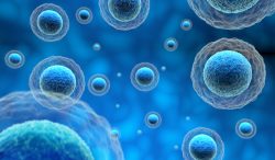 It was once thought that neurons and possibly heart cells, were the oldest cells in the human body. However, researchers have discovered that the mouse liver, brain and pancreas contain populations of proteins and cells with very long lifespans with some as old as neurons.
The findings at the Salk Institute demonstrating age mosaicism could be applied to nearly any body tissue for providing valuable information about the lifelong function of non dividing cells and how cells lose control over the integrity and control of proteins and important cell structures during the aging process.
The team was very surprised to discover cellular structures that are as old as the organism where they reside. This discovery suggests even greater cellular complexity than was previously imagined. It provides intriguing implications about how we think about the aging process of organs such as the heart, pancreas and brain.
Most brain neurons do not divide in adulthood and therefore experience a long lifespan and then age related decline. However, due to technical limitations, the life of cells outside the brain has been difficult to determine. Researchers have wondered how old the cells are in an organism.
There has been the general idea that neurons are old yet other cells in the human body are relatively young and will regenerate throughout the lifetime of the organism. The research team proceeded to see if there was the possibility that certain organs also have cells that are as long lived as brain neurons.
The researchers knew that most neurons will not be replaced during lifespan. They utilized them as an age baseline for comparison to other non dividing cells. They combined electron isotope labeling with a hybrid imaging method so they could visualize and then quantify protein and cell age and turnover in the liver, pancreas and brain in both old and young rodents.
To validate the method, the team initially determined the age of the neurons and found that they were as old as the organism. However, the cells that line blood vessels, known as endothelial cells, were also as old as the neurons. This means that some of the non neuronal cells do not replace or replicate themselves during lifespan.
The pancreas which is an organ whose function it is to maintain blood sugar levels and also secrete digestive enzymes, also showed cells at different ages. A small portion of the pancreas appeared as a puzzle of interconnected old and young cells. Some of the beta cells which release insulin replicated through lifetime and were relatively young. However, some did not divide and were long lived which is similar to neurons. Another type of cell called delta cells, did not divide at all. The pancreas is a great example of age mosaicism or a population of identical cells which are distinguished by their lifespans.
Previous research has suggested the liver has the ability to regenerate during adulthood so the team chose this organ with the expectation of seeing relatively young liver cells. However, the vast majority of liver cells in healthy adult mice were as old as the mice while cells which line blood vessels and stellate like cells, were much shorter lived. The liver also demonstrated ago moscaicism which indicates potential new paths of regenerative research of this organ.
Due to new visualizing technologies, the team was able to pinpoint the age of cells and their supra molecular complexes more precisely that before. This opens new paths for studying tissues, cells and organs in both normal and in diseased states.
By determining the age of sub cellular structures and cells in adult organisms, new insights into cell repair mechanisms and maintenance and the impact of cumulative changes during adulthood on development of diseases and health can occur.
The ultimate goal is to be able to utilize these mechanisms in an effort to delay and prevent age related decline of organs with limited cell renewal. The team plans to decipher the differences in lifespans for lipids and nucleic acids and also to understand how age mosaicism relates to diseases and overall health.
To view the original scientific study click here: Age Mosaicism across Multiple Scales in Adult Tissues.
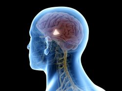

 Short bouts of exercise has been found to directly boost gene function which increases the connections between neurons in the hippocampus. This part of the brain is associated with memory and learning. Not only is exercise good for health, but according to recent research it can also help make you smarter!
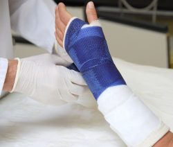 A team at the University of Illinois and the University of Pennsylvania created a new technique which uses flexible implantable bone stabilizing plates and stem cells to assist in faster healing of bone defects and large breaks. This new technique allows stem cells which are applied to break sites to experience a level of mechanical stress which they also do in developing embryos.
 A new discovery by scientists at the University of Nottingham indicates drinking a cup of coffee can actually stimulate brown fat. Brown fat is the body’s fat fighting defense which just might be the key to tackling diabetes and obesity.
 A new small scale controlled research trial, the first of its kind, has linked consumption of ultra processed foods to overeating and weight gain. People who eat these kinds of foods typically eat more calories when compared to eating a minimally processed diet.
 A simple tweak to a night owls sleeping patterns could help with sleep/wake timings, better eating habits, improved morning performance, and a decrease in stress and depression. Night owls are people with late waking and sleep habits.
 A new study conducted by researchers at the University of Bergen, Norway, have discovered a significant connection between good oral health and Alzheimer’s Disease. They determined that gingivitis has a very decisive role in whether someone develops this disease or not.
 It has been suspected that our body’s various circadian clocks are able to operate independently from our central clock located in the hypothalamus of the brain. At the University of California, Irvine, scientists have found a way to test the theory.