
How the Brain Affects Aging
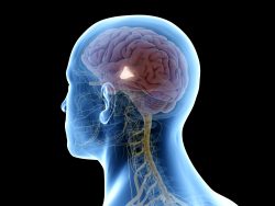
A new discovery by researchers at the Albert Einstein College of Medicine has shown that stem cells found in the brain’s hypothalamus affect how fast our body’s age. The discovery which was made in mice, could possibly lead to new strategies for defending against age related diseases and even extending lifespan.
The hypothalamus regulates critical processes including development, growth, metabolism and reproduction. Einstein researchers made the surprising discovery that it also regulates aging throughout the entire body. Now, they have pinpointed the exact cells in the hypothalamus that control the process of aging. These cells are a tiny population of adult neural stem cells which are known to be responsible for the formation of new brain neurons.
The research has shown that the number of hypothalamic neural stem cells will naturally decline throughout the life of an animal. This decline will accelerate with aging. The team also discovered that the effects of this stem cell loss are not irreversible. Through replenishing these stem cell or the molecules they produce, it is possible to slow down and even reverse a variety of the aspects associated with aging within the body.
To study whether stem cells found in the hypothalamus hold the key to aging, the team first looked at the fate of the stem cells as healthy mice aged. The number of these stem cells did begin to decline when the mice reached almost 10 months of age which is quit a few months before the normal signs of aging start to appear. By old age which is about two years in mice, most of these stem cells were gone.
The team then wanted to learn whether the progressive loss of these stem cells was actually causing the aging and was not only associated with it. They observed what happened after they selectively disrupted these stem cells in the hypothalamus in middle aged mice. The disruption greatly accelerated the aging compared with control mice. The mice with the disrupted stem cells also died earlier than what would be normal.
The question became, could adding stem cells to the hypothalamus actually counteract aging? The team sought to answer that question by injecting hypothalamic stem cells in the brains of the middle aged mice whose stem cells were destroyed along with injecting the brains of the normal older mice. In both groups of mice the treatment either slowed or reserved a variety of measures of aging.
The team found that the hypothalamic stem cells appeared to exert their anti aging effects by the release of molecules called microRNAs. Rather than being involved in the synthesis of protein, they instead played key roles in regulating gene expression. MicroRNAs are contained inside tiny particles known as exosomes which stem cells in the hypothalamus release into the cerebrospinal fluid of mice.
The team then extracted microRNA containing exosomes from the hypothalamic stem cells and injected them into the cerebrospinal fluid of two different groups of mice…middle aged mice whose hypothalamic stem cells were destroyed and the normal middle aged mice. The treatment showed significant slowing of aging in both groups of mice through a measure of tissue analysis and also behavioral testing which involved assessing any changes in the mice’s coordination, muscle endurance, cognitive ability and social behavior.
They are now working at identifying the particular populations of the microRNAs and even other factors secreted by these stem cells which are responsible for the anti aging effects. This is potentially a first step to possibly slowing down the aging process and also treating a variety of age related diseases.
To view the original scientific study click below
Hypothalamic stem cells control ageing speed partly through exosomal miRNAs
 Short bouts of exercise has been found to directly boost gene function which increases the connections between neurons in the hippocampus. This part of the brain is associated with memory and learning. Not only is exercise good for health, but according to recent research it can also help make you smarter!
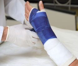 A team at the University of Illinois and the University of Pennsylvania created a new technique which uses flexible implantable bone stabilizing plates and stem cells to assist in faster healing of bone defects and large breaks. This new technique allows stem cells which are applied to break sites to experience a level of mechanical stress which they also do in developing embryos.
 A new discovery by scientists at the University of Nottingham indicates drinking a cup of coffee can actually stimulate brown fat. Brown fat is the body’s fat fighting defense which just might be the key to tackling diabetes and obesity.
 A new small scale controlled research trial, the first of its kind, has linked consumption of ultra processed foods to overeating and weight gain. People who eat these kinds of foods typically eat more calories when compared to eating a minimally processed diet.
 A simple tweak to a night owls sleeping patterns could help with sleep/wake timings, better eating habits, improved morning performance, and a decrease in stress and depression. Night owls are people with late waking and sleep habits.
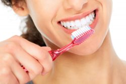 A new study conducted by researchers at the University of Bergen, Norway, have discovered a significant connection between good oral health and Alzheimer’s Disease. They determined that gingivitis has a very decisive role in whether someone develops this disease or not.
 It has been suspected that our body’s various circadian clocks are able to operate independently from our central clock located in the hypothalamus of the brain. At the University of California, Irvine, scientists have found a way to test the theory.
 New research has revealed that there are potential side effects and ongoing ramifications from long term protein intake or from certain types of amino acids. There has long been popularity and attention paid to consumption of proteins, however less attention has been paid to looking at its possible problems.