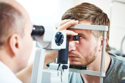
 Scientists at Harvard Medical School have successfully reversed age-related vision loss in mice through turning back the time on aged retina eye cells to bring back youthful functioning of the gene. This work is considered to be the first test of the possibility to reprogram, in a safe manner, complex tissues such as nerve cells found in the eye to an earlier age.
The team also has reversed loss of vision successfully in the mice that have a condition that mimics human glaucoma which is one of the leading causes of blindness globally.
The achievements represent the first successful attempts to reverse glaucoma induced loss of vision instead of merely stemming its progression. If future studies are replicated, this approach would show the way for therapies to promote repairing tissues of various organs and also reverse in humans aging and diseases that are age related.
The study can demonstrate that it is possible to reverse safely the age of complex tissues as the retina and restore its biological function to a youthful state. They caution that the findings need to be replicated in future studies, including in a variety of models of animals before human experiments. However, the results offer conceptual proof and also a pathway to designing treatments for a variety of diseases that are age-related.
If realized through future studies, the findings will possibly be transformative for the treatment of diseases that are age related such as glaucoma and also to the fields of medical therapeutics and biology for disease at large.
For the work they used an adeno-associated virus (AAV) as the vehicle for delivering three youth restoring genes into the retinas in mice – Oct4, Sox2, and Klf4 which during embryonic development are switched on.
The treatment showed multiple effects that were beneficial on the eyes of the mice. It firstly promoted regeneration of the nerve after damage to the optic nerves in mice. It also reversed vision loss in mice that had a condition that mimicked human glaucoma. And then it also reversed loss of vision in animals aging without glaucoma.
The new theory the research team used was based on why humans age. Almost all cells in a humans body have the same DNA molecules with dunctions that are widely diverse. To achieve the specialization of this degree, these cells can only read genes that are type specific. This regulatory function is the purview of the epigenome which is a system of specific patterns turning genes on and off without any alteration of the basic underlying DNA sequence of the gene.
The theory proposes that changes to the epigenome with time causes cells to read genes that are wrong and malfunction which promotes rise to diseases associated with aging. In the current study, the team hypothesizes that if DNA methylation, a way by which methyl groups are added onto DNA, does control aging, then deleting some of its footprints may reverse cell aging inside living organisms and eventually restore them to a more youthful and earlier state. Earlier work has achieved this in cells grown in lab dishes, thus falling short of showing the effect in a living organism. The new findings have demonstrated that the approach could be used in animals as well.
For the current study, the team wanted to use cells in the bodies central nervous system due to the fact that it is the first part of the body that is affected by aging. Following birth, the central nervous is prone to regeneration quickly.
In order to test if the young animals have the capacity to regenerate could be imparted to adult mice, the team had delivery of the modified three-gene combination per an AAV into retinal ganglion cells of adult mice who had optic nerve injury. The treatment resulted in a increase two-fold in the number of retinal ganglion cells surviving following the injury and increase five-fold in nerve regrowth.
At the start of the project, many of the team’s colleagues thought their approval would either be too dangerous or would fail to ever be used. However, their results promote this method is indeed safe and could possibly revolutionize eye treatments along with many other organs that are age affected.
Following the positive findings in mice with injuries of the optic nerve, the team partnered with other colleagues to find out if the three-gene cocktail could restore loss of vision due to glaucoma and also another test to see if the approach could reverse loss of vision stemming from the normal aging process.
In a mouse model that had glaucoma, the treatment led to increased nerve cell electrical activity and a notable increase in visual acuity. Remarkably, it did so following the glaucoma-induced vision loss had already occurred.
Scientists have rarely been successful in regaining visual function following an injury. This new approach which would successfully reverse multiple causes of loss vision in mice without the need for a retinal transplant, is basically a new treatment modality in regenerative medicine. The treatment also worked well in older, year old mice with vision diminishing due to normal aging.
The findings are encouraging and researchers believe that if these confirmed findings in further animal study, they may initiate clinical trials before two years to test the efficacy of this approach in people who have glaucoma.
To view the original scientific study click below
Reprogramming to recover youthful epigenetic information and restore vision.

 A University of Otago, New Zealand, study has found that three pillars of health which are exercising, quality of sleep and eating raw vegetables and fruit, promotes better health mentally and overall well-being feeling in younger adults. And the research found that the strongest predictor was better quality of sleep than sleep quantity.
 Plastics can have and impart to a human a variety of dangerous chemicals including endocrine disrupting chemicals (EDCs) that pose a threat to human health. A new report has reported the dangerous health effects of contamination that is widespread from the EDCs in plastics.
 According to new research at University Hospital A Coruna Spain, the time required to climb four flights of stairs provides an excellent indicator of heart health. Cardiologists use climbing stairs in a physical exam for that purpose.
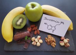 It is well known that intestinal health has a close link with healthy functioning of the brain. Researchers from the University of Tsukuba in Japan are now suggesting that normal sleep patterns may be influenced by gut bacteria through the ability to help create important chemical messengers in the brain such as dopamine and serotonin.
 A new study from the University of Warwick and University Hospitals Coventry and Warwickshire NHS Trust, has shown that your age is not a barrier to successful weight loss. People who are obese aged 60 and over can lose an the dame amount of weight as someone younger using only changes in their lifestyle.
  Evidence for the harmful effects of alcohol on brain health has been compelling. And now researchers in the UK and Australia have shown evidence suggesting three periods of dynamic changes in the brain that could be particularly sensitive to the harmful effects of alcohol – gestation (conception to birth), later adolescence (15 to 19 years of age) and older adulthood (over 65 years).
 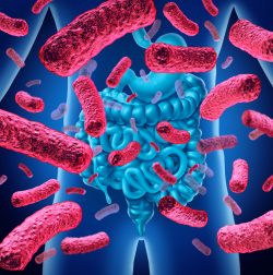 A new study has found links between gut bacteria and the active form of Vitamin D. This study has suggested that gut bacteria may play a vital role in converting inactive Vitamin D into its active, health benefiting form. Vitamin D is essential for maintaining healthy teeth and bones and for a strong immune system. The study has revealed a new understanding of Vitamin D and how it is typically measured.
 A study led by Massachusetts General Hospital (MGH) has found that even physical exercise in short bursts can create changes in the body’s levels of metabolites that can correlate to and might help gauge a person’s cardiovascular, cardiometabolic and long term health. The study shows how a time of 12 minutes consisting of acute cardiopulmonary exercise can impact more than 80% of circulating metabolites and includes pathways associated to a wide range of beneficial health outcomes.
  A recent study led by Dana King, a professor and chair of the Department of Family Medicine, along with research partner Jun Xiang, have found that glucosamine supplements may reduce overall mortality almost as well as regular exercise does. The new epidemiological study has provided encouraging results that are good indicators supporting their analysis.