
Excessive Training can make your Brain Tired
 Excessive training can make the body tired, however can it make the brain tired? According to a new study, the answer is yes!
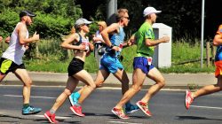
Researchers put triathletes on an excessive training load. They found they showed a form of mental fatigue. The fatigue showed reduced activity in the portion of the brain that is important for decision making. The athletes also showed more impulsive behavior, choosing immediate rewards over bigger ones that would take longer to achieve.
The lateral prefrontal portion of the brain that was affected by the overload in sport training was exactly the same which had been shown vulnerable to excessive work in earlier studies. This particular area of the brain appeared as the weak spot of the brain network which is responsible for cognitive control.
The two studies suggest a connection between physical and mental effort both of which require cognitive control. The reason for this essential control in demanding athletic training is that to maintain physical effort and to reach a distant goal, cognitive control is required. A person needs to control the automatic process that will make them stop when joints or muscles hurt.
The study originated from the National Institute of Sport, Expertise, and Performance in France which trains athletes for Olympic games. Some of the athletes suffered from over training syndrome. Their performance plummeted when they experienced an overwhelming sense of fatigue.
The question became, does this over training syndrome arise in part from neural fatigue in the brain which is the same kind of fatigue that can be caused by excessive intellectual work?
To discover the answer, the research team recruited 37 competitive male endurance athletes who had an average age of 35. The participants were assigned to either continue on with their normal training or to increase their training by 40% per session over a 3 week period.
The physical performance of the athletes was monitored during cycling exercises which were performed on rest days. The research team assessed their subjective experience of fatigue through questionnaires every 2 days. Behavioral testing and functional magnetic resonance imaging scanning experiments were also conducted.
The results showed that physical training overload caused the athletes to feel more fatigued. Additionally, they acted more impulsively in standard tests which were used to evaluate how they would make economic choices.
The tendencies were shown as a bias in favoring immediate rewards over delayed ones. The brains of the athletes who had been overloaded physically also indicated diminished activation of the lateral prefrontal cortex which is a key region of the executive control system when they made the economic choices.
The studies findings indicate that while endurance sport is generally good for health, overdoing it can lead to adverse effects on the brain. The findings bring attention to the fact that neural states matter. People don’t make the same decisions when the brain is in a state of fatigue.
The findings may be important not only for producing the best athletes, but also for economic choice theory which typically will ignore fluctuations in the neural machinery which is responsible for decision making. It also suggests it may be important to monitor fatigue levels in order to prevent bad decision making in the judicial, political and economic domains.
Future studies will explore why exerting control during sports training or intellectual work causes the cognitive control system harder to activate in subsequent tasks. The hope is to find strategies or treatments that can help prevent such neural fatigue and the following consequences.
To view the original scientific study click below
Neuro-computational impact of physical training overload on economic decision-making.
 Air pollution may have a harmful effect on more than just the heart and lungs. A new study has shown that it could also be making us less intelligent. The study conducted on elderly people living in China, has found that long term exposure to pollution in the air may hinder cognitive performance in both math and verbal tests.
  As we get older, it can sometimes become more difficult to keep our weight in check. New research at the Karolinska Institutet in Sweden along with collaboration with researchers at Uppsala University in Sweden and the University of Lyon in France, has discovered why this is.
  Biologists at MIT’s Koch Institute of Integrative Cancer Research have shown that high levels of ketone bodies which are molecules produced through the breakdown of fat, help our intestines maintain a large pool of adult stem cells. These stem cells are critical for keeping the intestinal lining in our bodies healthy.
 A researcher at the University of Guelph has discovered the gecko’s spinal cord tail houses a special cell type known as the radial glia. When the tail detaches, these cell types jump into action by making and proliferating different types of proteins in response to the injury. It is believed this discovery may have implications for spinal cord treatments in humans.
 Two new studies have explored the relationship between cognitive decline and oral health and also perceived social support and stress among older Chinese Americans. Two Rutgers studies found evidence that there is a key relationship between poor oral health and cognitive aspects such as executive function and memory.
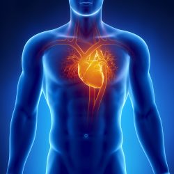 New research at the University of Calgary has discovered what was once an unidentified cell population in the pericardial fluid which is found inside the sac that surrounds the heart. This discovery could possibly lead to new treatments for people with injured hearts. The study was funded by the Heart and Stroke Foundation of Canada.
 A new study conducted at the University of California, Irvine, has found that online brain game exercises can help people in their 70’s and 80’s multitask cognitively even as well as people 50 years younger! This valuable tool, gives older adults the ability to handle today’s daily onslaught of information which can be very taxing for seniors.
 According to a recent study, 20 years of a sedentary lifestyle is linked to two times the risk of premature death when compared to being physically active. The HUNT study’s findings suggest that in order to get maximum health benefits from physical activity to protect against premature cardiovascular and all cause death, a person needs to be continually physically active.
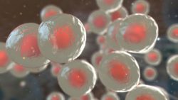 A research team at the Osaka University has conducted the world’s first corneal tissue transplant using reprogrammed stem cells derived from skin tissue. The patient was a Japanese woman in her 40’s who suffered from an epithelial stem cell deficiency in her cornea. This condition can make vision blurry and can lead to blindness.