How Exercise Slows Down Aging

Cycling

Staying active helps keep the body young and healthy! And a lifetime of regular exercise can help defy the aging process! A study conducted by researchers at the University of Birmingham and King?s College London, found that older people who had exercised all of their lives had the immunity, muscle mass and cholesterol levels of a young person.

The study included 125 amateur cyclists aged 55 to 79 ? 41 were female and 84 were male. The women had to be able to cycle 60 km in 5.5 hours and the men 100 km in under 6.5 hours. Anyone with high blood pressure or other health conditions, smokers and heavy drinkers were excluded.

The group underwent a variety of tests in a laboratory. The participants were compared to a group of adults who did not partake in regular physical activity. This group consisted of 75 healthy adults aged 57 to 80 and 55 healthy younger adults aged 20 to 36. The study showed that the active adults who exercised regularly did not show loss of muscle mass and strength. The cyclists also did not increase body fat or cholesterol levels with age and interestingly the men?s testosterone levels remained high indicating they may have avoided most of male menopause. The cyclists also had an immune system that did not seem to age either. The researchers also discovered that the thymus which makes immune cells called T cells and shrinks from the age of 20 and makes less T cells, was as active in the cyclists as those of a young person.

The research shows there is strong evidence that getting people to commit to regular exercise throughout their lives is a viable solution to living a longer, healthier life. Findings show that less than half of people over 65 do not exercise enough to remain healthy. And per Professor Janet Lord, Director of the Institute of Inflammation and Aging at the University of Birmingham, the findings debunk the assumption that aging means frailty. The studies findings indicate that the cyclists do not exercise because they are healthy, but because they had been exercising for a large portion of their lives, they are healthy. The exercise has allowed their bodies to age optimally, free from a lot of the problems caused by inactivity. Remove their activity, and the researchers agree that their health would most likely begin to decline.

Nearly everyone can exercise. It is a matter of finding an exercise that suits a person and can make a habit of it. It may lead to a later life that is healthier and productive! Hippocrates in 400 BC said ?Exercise is man?s best medicine?. Unfortunately, his message has been lost over time and we have become an increasingly sedentary society.

Reference: Ross D. Pollock, Katie A. O’Brien, Lorna J. Daniels, Kathrine B. Nielsen, Anthea Rowlerson, Niharika A. Duggal, Norman R. Lazarus, Janet M. Lord, Andrew Philp, Stephen D. R. Harridge. Properties of the vastus lateralis muscle in relation to age and physiological function in master cyclists aged 55-79 years. Aging Cell, 2018; e12735 DOI: 10.1111/acel.12735

Low-Fat vs. Low-Carbohydrate Diet: Which is Better?

fruit and vegetables

A new study has shown that it isn?t so much the type of diet, Low-Fat vs. Low Carb, but a more a matter of quality of the diet! A Stanford University study published in the Journal of the American Medical Association, has concluded that both diets can work equally well at shedding unwanted pounds as long as people consume healthy vegetables and whole foods and cutting back on refined grains, sugar and highly processed foods. And of course people have to stick to the diet!

Christopher Gardner, Director of Nutrition Studies at the Stanford Prevention Research Center, states they found no evidence that a person?s genotype (genetic makeup) or the individual way their body responds to insulin determines which diet type most likely to help them with their weight loss. Past research has indicated that genetic and insulin levels might explain why some people lost more weight on a low-carb diet vs. a low-fat diet or vice versa. The Stanford colleagues decided to study those assumptions by conducting a randomized clinical trial which is considered the gold standard for testing hypothesis.

The researchers recruited 609 adults aged 18 to 50 and evenly divided between men and women. All had a body mass index of between 28 and 40. All underwent a variety of tests at the beginning of the study including one test that looked for genetic variants linked to the metabolism of fasts and carbohydrates and one that measured the body?s response to insulin.

Participants were randomly assigned to either a low-carb diet or a low-fat diet which they were to follow for one year. Those on the low-fat diet were encouraged to cut down to 20 grams of fat per day for the first eight weeks of the study. Those in the low-carb group were asked to cut down to 20 grams of carbs per day. These were fairly large changes from what they had a their baselines.

During the first two months they were told they could add back small, incremental amount of carbs and fats until they reached a level they felt they could successfully maintain for the rest of their lives. At the end of the year study, those on the low-fat diet averaged 57 grams of fat daily and those on the low-carb diet were averaging 132 grams of carbohydrates daily which is much lower than the 83 grams of fat and 247 grams of carbs the groups were averaging at the beginning of the study.

During the study the groups were counseled on how to eat a healthy low-carb or low-fat diet. The main point was eating quality including buying fresh food and produce at the Farmer?s Market, cooking more yourself, eating with others, and watching snacking habits. They were also told to eat very little refined grains and sugar and focus on eating a lot of vegetables. And they were told to eat so that they didn?t feel deprived or hungry.

The main findings showed that the weight loss at the end of the year for both groups was very similar ? an average of 11 pound loss for the low-fat group and an average of just over 13 pounds for the low-carb group. The data also showed that people with genetic variations linked to a low-fat diet response were not more likely to lose weight on a low-fat diet nor were people with genetic variations linked to a low-carb diet response were likely to lose weight on a low-carb diet. Additionally, those with a poor insulin response were not more likely to lose weight on a low-carb diet vs. a low-fat diet. This proved the researchers original hypotheses true.

The study did have some limits. The participants tended to have higher levels of education that the general public and resources that enabled them access to high quality food. And the participants were only followed for one year. The studies findings do offer the best evidence to date for how low-fat and low-carb diets compare when it comes to weight loss and genetic factors.

Reference: Gardner CD, Trepanowski JF, Del Gobbo LC, Hauser ME, Rigdon J, Ioannidis JPA, Desai M, King AC. Effect of Low-Fat vs Low-Carbohydrate Diet on 12-Month Weight Loss in Overweight Adults and the Association With Genotype Pattern or Insulin SecretionThe DIETFITS Randomized Clinical Trial. JAMA. 2018;319(7):667?679. doi:10.1001/jama.2018.0245

Repairing Spinal Cord Injuries with Stem Cells

Spinal Cord

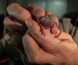
Led by researchers at University of California San Diego School of Medicine, a diverse team of neuroscientists and surgeons successfully grafted human neural progenitor cells into rhesus monkeys with spinal cord injuries. The grafts not only survived, but grew hundreds of thousands of human axons and synapses, resulting in improved forelimb function in the monkeys.

The findings, published online in the February 26 issue of Nature Medicine, represent a significant step in translating similar, earlier work in rodents closer to human clinical trials and a potential remedy for paralyzing spinal cord injuries in people.

“For more than three decades, spinal cord injury research has slowly moved toward the elusive goal of abundant, long-distance regeneration of injured axons, which is fundamental to any real restoration of physical function,” said Mark Tuszynski, MD, PhD, professor of neuroscience and director of the UC San Diego Translational Neuroscience Institute.

“While there was real progress in research using small animal models, there were also enormous uncertainties that we felt could only be addressed by progressing to models more like humans before we conduct trials with people,” Tuszynski said.

“We discovered, for example, that the grafting methods used with rodents didn’t work in larger, non-human primates. There were critical issues of scale, immunosuppression, timing and other features of methodology that had to be altered or invented. Had we attempted human transplantation without prior large animal testing, there would have been substantial risk of clinical trial failure, not because neural stem cells failed to reach their biological potential but because of things we did not know in terms of grafting and supporting the grafted cells.”

Gregoire Courtine, PhD, a professor and investigator at the Center for Neuroprosthetics and at the Brain Mind Institute, both part of the Swiss Federal Institute of Technology (EPFL) in Geneva, also conducts research seeking to restore function after spinal cord injury. He underscored the importance of the new findings.

“Dr. Tuszynski and his collaborators overcame a number of methodological difficulties specific to primates to obtain this breakthrough,” he said. “Direct translation of their work to humans would have failed, and yet too many studies are bypassing vital translational work in primate models that is necessary before human clinical trials.”

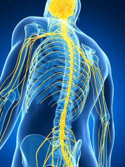
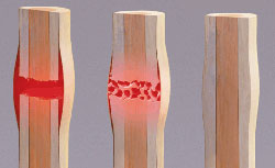
Successfully growing and proliferating functional grafted stem cells in spinal cord injuries is hindered by a multitude of innate, biological challenges. For example, the region surrounding the injury site — the so-called extracellular matrix inhibits growth in the same way that a superficial scar never resembles the original tissue in form or function. The injury site is abundant with inhibitory myelin proteins (used to make the insulating sheath around many nerve fibers) but lacks growth-promoting factors, such as neurotrophins, that would encourage regeneration of nerve cells’ axons and synapses.

Previous work by Tuszynski and others have found solutions or work-arounds for many of these obstacles, reporting notable progress using rodent models. The new work involves the use of human spinal cord-derived neural progenitor cells (NPCs) — stem cells destined to become nerve cells in the central nervous system (CNS) in rhesus monkeys, whose biology and physiology is much more similar to humans. Because the NPCs were derived from an 8-week-old human embryonic spinal cord, they possessed active growth programs that supported robust axon extension and appeared to be insensitive to inhibitors present in the adult CNS.

Two weeks after the initial injury (a period intended to represent the time required for an injured person to medically stabilize undergoing neural stem cell therapy), researchers grafted 20 million NPCs into the injury lesions in the monkeys, supported by a cocktail of growth factors and immune suppression drugs.

The work was done at the California National Primate Research Center at UC Davis. Most of the investigators are from UC campuses. “This highly complex translational project shows the value of collaborative research across UC campuses with unique facilities,” said co-author Michael Beattie, PhD, professor and director of research at the Brain and Spinal Injury Center at UC San Francisco.

Over the next nine months, the grafts grew, expressing key neural markers and sending hundreds of thousands of axons — the fibers through which nerve cells conduct signals to other nerve cells through the injury site to undamaged cells and tissue on the other side. Several months into the study, researchers noted that the monkeys began to display partial recovery of movement in their affected forelimbs.

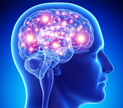
Notably, the team documented regeneration of corticospinal axons, which are essential for voluntary movement in humans, into the lesion sites the first such known documentation in a primate model.

Courtine at EPFL, who was not involved in the study, said the findings challenge decades of work on the mechanisms of regeneration failure and “definitely represent a landmark in regeneration medicine.” Nonetheless, he noted that the degree of functional improvement remained limited. “It is not surprising given that the functional integration of new cells and connections into the operation of the nervous system would require time and specific rehabilitation procedures,” he said.

“It’s possible that given a longer period of observation, greater recovery may have occurred,” said the study’s first author, Ephron S. Rosenzweig, PhD, an assistant adjunct professor in Tuszynski’s lab. “Axon regeneration, synapse formation, myelination these all take time, and are critical for neural function. Grafts, and the new circuitry they were part of, were still maturing at the end of our observations, so it seems possible that recovery might have continued.”

Tuszynski said work remains to be done before initiating human clinical trials, including production of a candidate neural stem cell line from humans that meets requirements of the Food and Drug Administration, and additional studies of safety. His group also continues to explore ways to further enhance the growth, distance and functionality of the regenerated cells.

“We seem to have overcome some major barriers, including the inhibitory nature of adult myelin against axon growth,” he said. “Our work has taught us that stem cells will take a long time to mature after transplantation to an injury site, and that patience will be required when moving to humans. Still, the growth we observe from these cells is remarkable — and unlike anything I thought possible even ten years ago. There is clearly significant potential here that we hope will benefit humans with spinal cord injury.”

Reference: Ephron S Rosenzweig, John H Brock, Paul Lu, Hiromi Kumamaru, Ernesto A Salegio, Ken Kadoya, Janet L Weber, Justine J Liang, Rod Moseanko, Stephanie Hawbecker, J Russell Huie, Leif A Havton, Yvette S Nout-Lomas, Adam R Ferguson, Michael S Beattie, Jacqueline C Bresnahan, Mark H Tuszynski. Restorative effects of human neural stem cell grafts on the primate spinal cord. Nature Medicine, 2018; DOI: 10.1038/nm.4502

Abstract: “We grafted human spinal cord?derived neural progenitor cells (NPCs) into sites of cervical spinal cord injury in rhesus monkeys (Macaca mulatta). Under three-drug immunosuppression, grafts survived at least 9 months post injury and expressed both neuronal and glial markers. Monkey axons regenerated into grafts and formed synapses. Hundreds of thousands of human axons extended out from grafts through monkey white matter and synapsed in distal gray matter. Grafts gradually matured over 9 months and improved forelimb function beginning several months after grafting. These findings in a ‘preclinical trial’ support translation of NPC graft therapy to humans with the objective of reconstituting both a neuronal and glial milieu in the site of spinal cord injury.”

Super-Agers Amazing Brain Health

Brain

A Super-aging Study being led by neuroscientist Emily Rogalski at Chicago?s Northwestern University may give new insight into how some people in the 80?s and 90?s keep the same sharp memory as someone else several decades younger. The work is a turn on the disappointing hunt for new drugs to help or prevent a variety of dementia related diseases as humans age.

The study team has conducted a battery of tests on more than 1,000 people who were thought to qualify and about 5 percent passed. The key memory challenge was for the participants to listen to 15 unrelated words and then a half-hour later recall at least nine of them which is the norm for 50-year-olds. The average 80-year old recalls five super-agers remember them all. Super-agers tend to be extroverts and have strong social networks, otherwise they come from all walks of life. Some were college graduates, some weren?t. Some have very high IQ?s while others are average. Some participants had experienced enormous trauma. Some were fitness buffs while others smoked. Some enjoyed alcohol while others were teetotalers. The differences made it difficult for the researchers to find a common trait for brain health.

Parts of the brain do shrink as people age. But it is deep within the brain that the researchers found compelling evidence that super-agers brains are more resilient against the ravages of aging. It turns out that super-agers brains do not shrink nearly as fast. Autopsies performed on some of the first super-agers in the study to die, show their brains harbor a lot more of a special kind of nerve cell in a deep brain region. Brain scans also showed that a super-agers cortex, an outer brain layer critical to memory and other key functions, is much thicker than normal for the individual?s age. It looks more like the cortex of a healthy 50 or 60 year old person?s brain. That could be related to what they were born with, but the researchers also say another clue is that their cortex just doesn?t shrink as fast over 18 months average 80 year olds experienced more than twice the rate of loss compared to super-agers.

Researchers also discovered that deep in the brain of the super-agers the attention region is much larger. And inside, autopsies showed that region of the brain was packed with unusual large, spindly von Economo meurons special and little understood neurons thought to play a role in social processing and awareness. The super-agers had four to five times more of those types of neurons compared to the typical octogenarian and even more than the average young adult.

The super-agers are living long and living well. Rogalski wonders?are there modifiable things people can do today in everyday lives to do the same. The study is still looking for Super-ager clients who are in the 80?s and 90?s ? who are very active intellectually or physically who can contribute to the study. Another study being conducted at the University of California, Irvine, is also studying the old people in their 90?s and above. Some of these participants have Alzheimer’s, some have maintained excellent memories and some are in between. About 40 percent of the participants who had died, showed no symptoms of Alzheimer?s disease at death although their brains showed full-fledged signs of Alzheimer’s in their brains. Interestingly, the researchers found varying amounts of amyloid and tau, hallmark proteins in people with Alzheimer’s, in the brains of some of these super=agers which makes the researchers wonder how these people deflect damage perhaps they have different pathways to brain health.

Gut Microbiota Study Brings New Hope for Improving Health

Microbiome

Our microbiome is the personal complement of mostly friendly bacteria we carry around with us. Study after study has found that our microbiome affects nearly every aspect of our health; and its microbial composition, which varies from individual to individual, may hold the key to everything from weight gain to moods. Some microbiome researchers had suggested that this variation begins with differences in our genes; but a large-scale study conducted at the Weizmann Institute of Science challenges this idea and provides evidence that the connection between microbiome and health may be even more important than we thought.

Indeed, the working hypothesis has been that genetics plays a major role in determining microbiome variation among people. According to this view, our genes determine the environment our microbiome occupies, and each particular environment allows certain bacterial strains to thrive. However, the Weizmann researchers were surprised to discover that the host’s genetics play a very minor role in determining microbiome composition only accounting for about 2% of the variation between populations.

The research was led by research students Daphna Rothschild, Dr. Omer Weissbrod and Dr. Elad Barkan from the lab of Prof. Eran Segal of the Computer Science and Applied Mathematics Department, together with members of Prof. Eran Elinav’s group of the Immunology Department, all at the Weizmann Institute of Science. Their findings, which were recently published in Nature, were based on a unique database of around 1,000 Israelis who had participated in a longitudinal study of personalized nutrition. Israel has a highly diverse population, which presents an ideal experimental setting for investigating the effects of genetic differences. In addition to genetic data and microbiome composition, the information collected for each study participant included dietary habits, lifestyle, medications and additional measurements. The scientists analyzing this data concluded that diet and lifestyle are by far the most dominant factors shaping our microbiome composition.

If microbiome populations are not shaped by our genetics, how do they nonetheless interact with our genes to modify our health? The scientists investigated the connections between microbiome and the measurements in the database of cholesterol, weight, blood glucose levels, and other clinical parameters. The study results were very surprising: For most of these clinical measures, the association with bacterial genomes was at least as strong, and in some cases stronger, than the association with the host’s human genome.

According to the scientists, these findings provide solid evidence that understanding the factors that shape our microbiome may be key to understanding and treating many common health problems.

In Tampa, Florida a group of doctors have already discovered how to permanently transplant the gut microbiota from a healthy person to someone with various health challenges and poor microbiota. This is being done, however currently is limited to only certain serious conditions.

According to Segal “We cannot change our genes, but we now know that we can affect and even reshape the composition of the different kinds of bacteria we host in our bodies. So the findings of our research are quite hopeful; they suggest that our microbiome could be a powerful means for improving our health.”

The field of microbiome research is relatively young; the database of 1,000 individuals collected at the Weizmann institute is one of the most extensive in the world. Segal and Elinav believe that over time, with the further addition of data to their study and those of others, these recent findings may be further validated, and the connection between our microbiome, our genetics and our health will become clearer.

Reference: Daphna Rothschild, Omer Weissbrod, Elad Barkan, Alexander Kurilshikov, Tal Korem, David Zeevi, Paul I. Costea, Anastasia Godneva, Iris N. Kalka, Noam Bar, Smadar Shilo, Dar Lador, Arnau Vich Vila, Niv Zmora, Meirav Pevsner-Fischer, David Israeli, Noa Kosower, Gal Malka, Bat Chen Wolf, Tali Avnit-Sagi, Maya Lotan-Pompan, Adina Weinberger, Zamir Halpern, Shai Carmi, Jingyuan Fu, Cisca Wijmenga, Alexandra Zhernakova, Eran Elinav, Eran Segal. Environment dominates over host genetics in shaping human gut microbiota. Nature, 2018; DOI: 10.1038/nature25973

Stem Cell and Gene Therapy Utilized to Repair Severe Bone Fractures

Bone Fracture

A Study led by a team of researchers at Cedars-Sinai has completed a study that may revolutionize orthopedics. They have successfully repaired severe limb fractures in laboratory animals. They used an innovative technique that signaled bones to regrow its own tissue combining an engineering approach with a biological approach for advancing regenerative engineering. Six different departments at Cedar-Sinai and researchers from Hebrew University, University of Rochester and University of California were involved in the study further demonstrating the effectiveness of utilizing a variety of diverse disciplines to further solutions to today?s medical challenges.

Currently more than 2 million bone grafts are performed worldwide each year due to severe injuries such as traffic accidents, war or major surgery. These injuries many times create a large gap between the edges of a fracture resulting in the inability of the bone to bridge the gap on its own. Currently pieces from a patient?s or a donor?s bone must be implanted into the gap. These bone grafts unfortunately carry disadvantages in skeletal repair. Many times healthy bones are not available for the repairs and implants that do occur can prolong pain and longer hospital stays. And some repairs fail.

The researchers devised a new technique as a possible alternative to the bone grafts. They constructed a matrix of collagen which they implanted in the space between two sides of a fractured leg bone in the animals. Collagen is a protein that the body uses to build bones. The matrix recruited the broken leg?s own stem cells into the gap over two weeks. The team delivered a bone-inducing gene directly into the stem cells to being the bone repair process. The process involved using an ultrasound pulse and micro-bubbles which facilitated the gene entry into the cells. Just eight weeks after the surgery, the bone gaps were closed and the fractured legs were healed in all lab animals used in the treatment process. Furthermore, the bone growth that occurred in the gap was as strong as surgical bone grafts.

By demonstrating that gene delivery into an animal?s own stem cells through ultra-sound was effective, the study has opened up new possibilities for treating non-healing bone fractures.

Reference: Maxim Bez1, Dmitriy Sheyn, Wafa Tawackoli, Pablo Avalos, Galina Shapiro1, Joseph C. Giaconi, Xiaoyu Da, Shiran Ben David, Jayne Gavrity, Hani A. Awad, Hyun W. Bae, Eric J. Ley, Thomas J. Kremen, Zulma Gazit, Katherine W. Ferrara, Gadi Pelled, Dan Gazit; In situ bone tissue engineering via ultrasound-mediated gene delivery to endogenous progenitor cells in mini-pigs; Science Translational Medicine, 17 May 2017: Vol. 9, Issue 390, eaal3128 DOI: 10.1126/scitranslmed.aal3128

Abstract: More than 2 million bone-grafting procedures are performed each year using autografts or allografts. However, both options carry disadvantages, and there remains a clear medical need for the development of new therapies for massive bone loss and fracture nonunions. We hypothesized that localized ultrasound-mediated, microbubble-enhanced therapeutic gene delivery to endogenous stem cells would induce efficient bone regeneration and fracture repair. To test this hypothesis, we surgically created a critical-sized bone fracture in the tibiae of Yucat?n mini-pigs, a clinically relevant large animal model. A collagen scaffold was implanted in the fracture to facilitate recruitment of endogenous mesenchymal stem/progenitor cells (MSCs) into the fracture site. Two weeks later, transcutaneous ultrasound-mediated reporter gene delivery successfully transfected 40% of cells at the fracture site, and flow cytometry showed that 80% of the transfected cells expressed MSC markers. Human bone morphogenetic protein-6 (BMP-6) plasmid DNA was delivered using ultrasound in the same animal model, leading to transient expression and secretion of BMP-6 localized to the fracture area. Micro?computed tomography and biomechanical analyses showed that ultrasound-mediated BMP-6 gene delivery led to complete radiographic and functional fracture healing in all animals 6 weeks after treatment, whereas nonunion was evident in control animals. Collectively, these findings demonstrate that ultrasound-mediated gene delivery to endogenous mesenchymal progenitor cells can effectively treat nonhealing bone fractures in large animals, thereby addressing a major orthopedic unmet need and offering new possibilities for clinical translation.

Why Do Naked Mole Rats Live Such Long Cancer Free Lives?

Naked Mole Rat

With their large buck teeth and wrinkled, hairless bodies, naked mole rats won’t be winning any awards for cutest rodent. But their long life span of up to 30 years is the longest of any rodent and their remarkable resistance to age-related diseases, offer scientists key clues to the mysteries of aging and cancer. Mice are about the same size, however they only live 2 or 3 years and often develop cancer.

That’s why University of Rochester biology professors Vera Gorbunova and Andrei Seluanov and postdoctoral associate Yang Zhao studied naked mole rats to see if the rodents exhibit an anticancer mechanism called cellular senescence and, if so, “how the mechanism might work differently than in short-lived animals, like mice,” says Zhao, the lead author of the study, published in PNAS.

Cellular senescence is an evolutionary adaptation that prevents damaged cells from dividing out of control and developing into full-blown cancer. However, senescence has a negative side too: by stopping cell division in order to prevent potential tumors, it also accelerates aging.

Previous studies indicated that when cells that had undergone senescence were removed from mice, the mice were less frail in advanced age as compared to mice that aged naturally with senescent cells intact.

Researchers therefore believed senescence held the key to the proverbial fountain of youth; removing senescent cells rejuvenated mice, so perhaps it could work with human beings. Companies began investigating drugs known as senolytic agents that would kill senescent cells and translate the anti-aging effects to humans.

But is eliminating senescence actually the key to preventing or reversing age-related diseases, namely cancer?

“In humans, as in mice, aging and cancer have competing interests,” Gorbunova says. “In order to prevent cancer, you need to stop cells from dividing. However, to prevent aging, you want to keep cells dividing in order to replenish tissues.”

Gorbunova and Seluanov have long researched cancer and its relation to aging and DNA repair. They have identified several mechanisms that contribute to longevity and cancer resistance in naked mole rats, including the chemical HMW-HA (high molecular weight hyaluronan). But they believe there are more pieces to the puzzle.

In their recent study, Zhao, Seluanov, Gorbunova, and their collaborators compared the senescence response of naked mole rats to that of mice, which live a tenth as long only about two to three years. “We wanted to look at these animals that pretty much don’t age and see if they also had senescent cells or if they evolved to get rid of cell senescence,” Seluanov says.

Their unexpected discovery? Naked mole rats do experience cellular senescence, yet they continue to live long, healthy lives; eliminating the senescence mechanism is not the key to their long life span. “It was surprising to us that despite its remarkable longevity the naked mole rat has cells that undergo senescence like mouse cells,” Gorbunova says.

The researchers found that although naked mole rats exhibited cellular senescence similar to mice, their senescent cells also displayed unique features that may contribute to their cancer resistance and longevity.

The cellular senescence mechanism permanently arrests a cell to prevent it from dividing, but the cell still continues to metabolize. The researchers found that naked mole rats are able to more strongly inhibit the metabolic process of the senescent cells, resulting in higher resistance to the damaging effects of senescence.

“In naked mole rats, senescent cells are better behaved,” Gorbunova says. “When you compare the signals from the mouse versus from the naked mole rat, all the genes in the mouse are a mess. In the naked mole rat, everything is more organized. The naked mole rat didn’t get rid of the senescence, but maybe it made it a bit more structured.”

Although their long life span is not a result of eliminating senescence, the more structured response to senescence allows them to have more systematic changes in their genes and have more orchestrated pathways being regulated. We believe this is beneficial for longevity and cancer resistance.”

Reference: Yang Zhao, Alexander Tyshkovskiy, Daniel Mu?oz-Esp?n, Xiao Tian, Manuel Serrano, Joao Pedro de Magalhaes, Eviatar Nevo, Vadim N. Gladyshev, Andrei Seluanov, Vera Gorbunova. Naked mole rats can undergo developmental, oncogene-induced and DNA damage-induced cellular senescence. Proceedings of the National Academy of Sciences, 2018; 201721160 DOI: 10.1073/pnas.1721160115

Why Does Short Term Memory Decline As We Get Older?

Memory

Most people suffer a decline in short term memory as they become older. It is common for instance for an elderly person to tell a friend or relative the same thing multiple times over a period of hours or days because they don’t remember they have already done so. Aging or impaired brains can once again form lasting memories if an enzyme that applies the brakes too hard on a key gene is lifted, according to University of California, Irvine neurobiologists.

“What we’ve discovered is that if we free up that DNA again, now the aging brain can form long-term memories normally,” said senior author Marcelo Wood, UCI’s Francisco J. Ayala Chair in Neurobiology & Behavior, who will present the findings at the American Association for the Advancement of Science’s annual meeting, in Austin, Texas. “In order to form a long-term memory, you have to turn specific genes on. In most young brains, that happens easily, but as we get older and our brain gets older, we have trouble with that.”

That’s because the 6 feet of DNA spooled tightly into every cell in our bodies has a harder time releasing itself as needed, he explained. Like many body parts, “it’s no longer as flexible as it used to be.” The stiffness in this case is due to a molecular brake pad called histone deacetylase 3, or HDAC3, that has become “overeager” in the aged brain and is compacting the material too hard, blocking the release of a gene called Period1. Removing HDAC3 restores flexibility and allows internal cell machinery to access Period1 to begin forming new memories.

Researchers had previously theorized that the loss of transcription and encoding functions in older brains was due to deteriorating core circadian clocks. But Wood and his team, notably postdoctoral fellow Janine Kwapis, found that the ability to create lasting memories was linked to a different process the overly aggressive enzyme blocking the release of Period1 in the same hippocampus region of the brain.

That’s potentially good news for developing treatments. “New drugs targeting HDAC3 could provide an exciting avenue to allow older people to improve memory formation,” Wood said.

Foods That Make You Smarter

Monosaturared Fatty Acid Foods

A study conducted at the University of Illinois has revealed that monounsaturated fatty acids (MUFAs) which are a class of nutrients found in nuts, avocados and olive oils are linked to general intelligence. Additionally they discovered that this relationship is driven by the correlation of MUFAs and the organization of the brain’s attention network. Researchers know that nutrition is linked to cognitive performance, however they had not previously pinpointed what underlies the connection.

The study published in the Nuerolmage Journal, involved 99 healthy older adults. It compared the patterns of fatty acid nutrients found in blood samples, functional MRI data which measured the efficiency of the brain networks and the results of a general intelligence test. The goal was to better understand how nutrition could be used to support cognitive performance and to study ways in which nutrition might also influence functional organization of the brain.

The researchers studied the relationship between groups of fatty acids and brain networks which underlie general intelligence. The goal was to understand whether the brain network organization mediated the relationship between general intelligence and fatty acids. Researchers were inspired by previous studies suggesting cognitive benefits of the Mediterranean diet which is naturally rich in MUFAs. Nutrients in the participants blood were examined and researchers found that the fatty acids clustered into saturated fatty acids and monounsaturated fatty acids. Rather than focusing on single nutrients, the researchers wanted to focus on broader dietary patterns.

The study revealed that general intelligence was associated with the brains dorsal attention network which plays a key role in everyday problem solving and attention-demanding tasks. The scientists found that general intelligence was associated with how efficiently the dorsal attention network is functionally organized using a measure called small-world propensity which describes how efficiently the neural network is connected within locally clustered regions as well as across globally integrated systems.

What they found was participants with higher levels of MUFAs in their blood had greater small-world propensity in their dorsal attention network. Taken together with an observed correlation between higher levels of MUFAs and greater general intelligence the findings suggest a pathway by which MUFAs affect cognition. The findings provide novel evidence that MUFAs are related to the dorsal attention network and how optimal this network is functionally organized. The results suggest that the dorsal attention network must be taken into consideration when understanding the relationship between MUFAs and general intelligence. It is part of the underlying mechanism that contributes to the relationship.

The hope is these findings will guide additional research into how nutrition affects intelligence and cognition. Further studies would involve an interventional study over time to see whether long-term MUFA intake influences brain network intelligence and organization. The evidence can certainly motivate promising new directions for future research in nutritional cognitive neuroscience.

Reference: Marta K. Zamroziewicz, M. Tanveer Talukdar, Chris E. Zwilling, Aron K. Barbey. Nutritional status, brain network organization, and general intelligence. NeuroImage, 2017; 161: 241 DOI: 10.1016/j.neuroimage.2017.08.043

Fast Food Continues to Harm Immune System Long After It’s Eaten

Fast Food
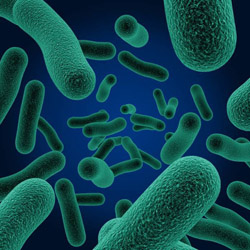
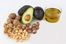
A recent study by a team at the Institute for Innate Immunity of the University of Bonn has discovered even more negative consequences as the result of a diet of fast food. Unhealthy food with a diet high in unhealthy fat and calories makes the body?s immune defenses aggressive even after switching to a healthy diet. The immune system reacts to this type of diet in a similar manner as it does to a bacterial infection. All of which leads to disease and other negative health ramifications.

The scientists put mice on a ?Western? Diet for one month. The diet was high in fat, high in sugar and low in fiber. The mice developed a strong inflammatory response throughout the body similar to symptoms with an infection with dangerous bacteria. The unhealthy diet resulted in an unexpected increase in the number of immune cells in the blood of the mice especially monocytes and granulocytes which help fight off bacteria, viruses and fungi. This indicated the involvement of immune cell progenitors in the bone marrow. To further understand these findings, bone marrow progenitors for major immune cell types were isolated from the mice fed a Western Diet and mice fed a healthy controlled diet. Genomic studies indicated the mice receiving the Western Diet did activate a large number of genes in the progenitor cells, genes responsible for proliferation and maturation. Findings indicate that fast food causes the body to very quickly recruit a powerful and huge army. When the mice were put back on their typical diet of cereal for another four weeks, the acute inflammation disappeared. However, what did not disappear was the genetic reprogramming of the immune cells and their precursors. Even after this length of time, the genes that had been turned on during the fast food diet were still active.

Only recently has it been discovered that the innate immune system has a form of memory. Following an infection, the body?s defenses remain in an alert state called ?innate immune training?. This allows the body?s defenses to respond more quickly to a new attack. In the mice, this process was triggered by an unhealthy diet rather than bacterium.

The scientists then examined the blood cells from 120 subjects. They identified the responsible ?fast food sensor? in immune cells. In some of the subjects, the innate immune system showed a strong training effect with researchers discovering genetic evidence of the involvement of a so-called inflammasomes. Inflammasomes are key intracellular signaling complexes which recognize infectious agents and other harmful substances which subsequently release highly inflammatory messages. In addition to the inflammatory response, this also has long-term consequences for the immune system?s responses. The Western diet changes the way in which genetic information is packaged.

This genetic material is stored in the DNA with each cell containing several DNA strands. They are typically wrapped around certain proteins in the nucleus and therefore many genes in the DNA cannot be read as they are too inaccessible. An unhealthy diet causes some of the normally hidden pieces of DNA to unwind and this area of the genetic material can be read much easier as long as the temporary unwrapping remains active. The inflammasomes trigger epigenetic changes and the immune system consequently reacts to even small stimuli with stronger inflammatory responses. This inflammatory response then accelerates the development of a variety of diseases and contributes to the growth of lipids and immune cells which will migrate to altered vessel walls which may result in serious health consequences.

These findings have important societal relevance. In recent centuries the average life expectancy has steadily increased in Western countries. That trend is currently being broken for the first time with individuals born today living an average shorter life than their parents. Too little exercise and unhealthy diets play a decisive role. The researchers strongly emphasize a healthy diet as the foundation for developing early habits that can affect long-term health.

Journal Reference: Anette Christ, Patrick G?nther, Mario A.R. Lauterbach, Peter Duewell, Debjani Biswas, Karin Pelka, Claus J. Scholz, Marije Oosting, Kristian Haendler, Kevin Ba?ler, Kathrin Klee, Jonas Schulte-Schrepping, Thomas Ulas, Simone J.C.F.M. Moorlag, Vinod Kumar, Min Hi Park, Leo A.B. Joosten, Laszlo A. Groh, Niels P. Riksen, Terje Espevik, Andreas Schlitzer, Yang Li, Michael L. Fitzgerald, Mihai G. Netea, Joachim L. Schultze, Eicke Latz. Western Diet Triggers NLRP3-Dependent Innate Immune Reprogramming. Cell, 2018; 172 (1-2): 162 DOI: 10.1016/j.cell.2017.12.013