
New Way to Generate Human Heart Cells from Stem Cells
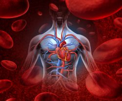 A new study is trying to understand early heart disease and development in relation to the mechanisms that determine cell fate. PSCs, or human pluripotent stem cells can conceivably produce a tissue the human body needs for repair. But developing technology for this to happen for a specialized cell, for example a beating heart muscle cell, has required intricate knowledge of developmental pathways and regulatory factors.
Although there has been important technological advances, types of cells that are obtained from human PSCs are still functionally immature. For example, in a human beating heart muscle cell (cardiomyocyte), the applications are limited in disease modeling and regenerative medicine. The cells that are generated don’t fully portray those that are found in the adult heart but are instead more similar to a fetal heart that is 20-weeks old.
The team utilized mouse embryo and cardiomyocytes to look into mechanisms that determine the fate of cells by modulating the peroxisome proliferator activated receptor (PPAR) signaling pathway. It plays a vital role in the development of cardiomyocytes to repeat in culture, and the cell in the womb to maturate and to progress to its specialization.
They were able to identify a response from isoform specific maturation, where PPARdelta signaling activation had the ability to augment the structural, metabolic and contractile maturation of hPSC-CMs. This being the first incident where PPAR signaling has been decoded in such an isoform specific way.
The specificity of PPARdelta, however, not PPARalha to show such an efficient effect on cardiac maturation was unexpected. The new strategy for maturation provided an easy and robust way to generate in culture mature heart cells. These can be used for a variety of applications such as disease modeling, drug screening or therapy for cell replacement in failing hearts.
The researchers were able to identify how the protein PPARdelta played a role by inducing a metabolic receptor from glycolysis to fatty acid oxidation in a heart muscle that was lab generated. This is a vital step in the controls and maturation no matter if these cells produce energy from fatty acids or glucose.
They showed that the signaling of PPARdelta plays an important role. It has the ability to turn on gene regulatory networks which increase the organization and number of mitochondria and peroxisomes, fatty acid oxidation, myofibril lay out, the contractility and the size of heart muscle cells.
To purify hPSC-CMs they are exposed to lactate and this treatment will trigger an independent molecular reaction for maturation of heart cells. In the study, the team exhibited lactate treatment along with activation of PPARdelta that increased further oxidative metabolism which allows energy generation from both fatty acids and carbohydrates.
The team developed a publicly and comprehensive accessible gene expression dataset of transcriptomic changes in hPSC-CMs which might be valuable to scientists studying PPAR lactate selection, PPAR signaling, or screening targets for drug or research testing.
The team is interested in analyzing disorders of fatty acid oxidation. This will depend on having CMs that primarily utilize fatty acid oxidation as a source of energy. They will investigate using the mature hPSC-CMs in transplants following an infarct.
The work will expand opportunities to further research biology of the human heart through multi-disciplinary avenues that incorporate transcriptomics, developmental biology, drug testing, and contractile measurements. They are headed to understanding how to use their knowledge of human development in order to improve access to human cell types that are mature.
To view the original scientific study click below:
PPARdelta activation induces metabolic and contractile maturation of human pluripotent stem cell-derived cardiomyocytes
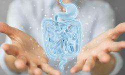 It is known that the intestinal lining needs to regenerate daily to be a powerful barrier to counter pathogens while allowing nutrients to be absorbed. The responsibility for this comes from the intestine’s stem cells. They need to meet a level of constant replenishment and repair. But, for this to happen, the stem cell needs to decide if the conditions of the intestine are receptive. If the stem cell makes the wrong decision or coordinates it poorly, intestinal cancer or diseases could occur.
 The indoor environment is usually more dangerous than the outdoors, with 90% of people’s time spent indoors. Chemicals from a variety of sources such as outdoor pollutants can seep into your home and cause problems for you there too! When it comes to health risks our bodies are constantly being bombarded by new chemicals – some good (like oxygen), others bad such as viruses or bacteria which want nothing better then an opportunity to live off of us. You might be surprised to learn that we are extremely harmful traveling emission sources of chemicals, including those found in our skin and breath.
 It has been found from reviewing 49 studies from 1800 weightlifters that increasing protein intake by double the amount increased strength by 9% and added about 1 lb of muscle growth. The participants worked out for 6 weeks and lifted weights at least twice per week.
  New research has identified how cells communicate to repair muscle damage. When a muscle is damaged, stem cells and immune cells work together to repair the damage. But how these cells interact to complete the removal process of dead tissue and make new muscle fibers had been a mystery. Scientists have discovered that hyaluranic acid is the essential molecule that contributes to this interaction.
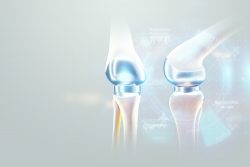 A new study has found a way to program stem cells in order to grow new cartilage on a 3-D template of the hip joint ball. This cartilage releases anti-inflammatory molecules that assist in fending off new arthritis occurrences. The new technology could supply an alternative to traditional hip replacement surgery and could remove the need for surgery for joint replacement surgery in some people.
  Hair follicle cells divide and die. But a new study has discovered a single chemical called TGF-beta that determines when this happens. It could ultimately treat baldness and may speed wound healing. Since follicles are a stem cell source they have the unique capability to be able to turn into other types of cells. This stem cell adaptability creates a path for repair of tissues and organs that have been damaged.
 While we sleep there are brain circuits and neuron learning connections that are active. They help us establish differences between items that are unrelated. The ability to remember indirect or arbitrary links between people, objects or events is called relational memory. This is what helps you put names with faces, find your keys or remember to turn off the stove before you leave the house.
 As we age, we lose muscle mass and the risk of dementia, heart disease, and immune function decreases. As the years go by, it becomes more difficult for humans to rebound from injury, a workout or illness. Aging takes a big toll on muscle tissue. Scientists have discovered that one type of activity in particular puts this process in reverse.
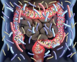 There are trillions of benign bacteria that live in our intestines. The immune system keeps them in constant balance which in turn ensures they are harmless to humans. A team has been able to demonstrate how some natural antibodies keep them in check. Their discoveries could make a substantial contribution to developing superior vaccines.