
How Eating Tomatoes Affects Gut Microbiome
 In a recent study, researchers found that eating a diet high in tomatoes over a two week period increased the diversity of gut microbes and altered bacteria toward an improved profile in young pigs. After observing the results with a short term intervention, the research team plans on moving into similar studies in people. The goal is that by looking at changes related specifically within the gut microbiome, we can see if any differences occur after consuming diets high or low in tomato content vs none at all.
Tomatoes are a popular vegetable, and research has shown that people who eat them tend to have less chronic health problems than those without. But what impact does consuming tomatoes really have on our guts? The new findings offer some insight into this mystery and show an avenue worth researching.
Microorganisms in our gut play an important role, but we still don’t fully understand their effects on human health. One way that research has tried to explore this question is by studying the link between dietary patterns and compositional differences among people’s guts by including food specific changes related specifically with certain foods like tomatoes. Ultimately they would like to see if there are any links between what type of bacteria someone gets through diet or other factors such as personal preference when it comes down how healthy they feel overall. The data shows us some interesting information about microbiome.
In this study, researchers compared the effects of feeding ten young pigs either an ordinary diet or one specially fine-tuned for them. The control group was given standard feed while those in the test group were fed food containing freeze dried tomatoes which are high in fiber and sugar but low on fat/calories content. They also took care not to interact too much with these animals during testing because it could potentially cause changes within their microbiome. Microbes in the pig’s guts were detected at both 7 and 14 days following their diets starting.
The study found that when pigs were fed a diet rich in tomatoes, their gut bacteria changed. The ratio between bacterial phyla Bacteroidetes to Bacillota, which has been linked with positive health outcomes, was higher during this time period compared to before when they were eating simpler foods such as grains or vegetables. Through their investigation, the researchers have characterized which microbes are present and how their relative abundance changed with tomato intervention.
The scientists want to learn more about how eating tomatoes changes our gut microbes and what this does for long-term health. They also aim on understanding the complex interplay between food intake, species diversity of bacteria in your body with additional research that could lead towards better dietary recommendations as well.
To view the original scientific study click below:
Short-Term Tomato Consumption Alters the Pig Gut Microbiome toward a More Favorable Profile
 In what could be a groundbreaking development, researchers have uncovered an immune response within the nose that normally works to fight off viruses implicated in upper respiratory illnesses. However, when temperatures dip below certain levels this protective mechanism is weakened leaving us more susceptible to infections such as colds and flu. The findings may explain why these diseases are much more common during colder weather conditions.
 You might be surprised to learn that a daily cup of tea could help you enjoy many years of good health in later life. If it’s not your preference, there are other things such as flavonoids found in some commonly consumed foods and beverages like green or black teas, nuts, apples, berries, citrus fruit, etc., all with the potential for long term benefits. New research shows they may actually do more than we thought.
 Brushing removes plaque and food debris, but the bristles of a toothbrush can’t reach deep in between teeth to remove it all. Flossing alone won’t be enough to keep you from getting gum disease and other oral complications because it only removes some of the plaque that has been collected on them throughout the day. In order for a person’s mouth to stay as clean as possible they need both an efficient routine with toothbrush and floss daily. As a result there’s less dental plaque which can lead to gum disease. But, should you brush or floss first?
 Electric lights have been a big change in the way we live our lives. It has been only about 130 years ago that they were invented bringing drastic changes to how humans function on a daily basis including our sleep patterns. For those who want better health or just more peace when it comes to sleeping at night make sure there isn’t any light coming into your bedroom. The dark will help you relax faster which leads towards a happier mood overall and improved physical health.
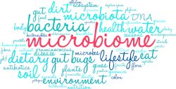 The blood metabolome is the small molecules found in your bloodstream that can interact with everything from brain function to bodily organs. We’re only just starting to understand the incredible impact our gut microbiome has on shaping molecules found in our blood. Figuring out what governs this variation could pave the way for precision approaches when it comes down to health and disease status.
 If you’re one of the millions of Americans who suffer from chronic constipation, relief may be closer than you think. According to a recent study, 16% of Americans have chronic constipation, and the odds rise to 33% for ages 60 and over. A recent study shows this unpleasant problem may have a pleasant solution.
  Intensive physical therapy and stem cell grafts together can boost the functionality of spinal cord injuries more than either treatment alone. Researchers found this in animal models, where tissue growth, repair, and functionality were increased following the combination of therapies. The stem cells promote growth and healing of the surrounding tissues, while the physical therapy helps to improve movement and function.
  If you’re looking to improve your muscular strength without the need for exercise, then this is good news! Green leafy vegetables are a great source of nitrates, and just one cup per day can provide a significant boost in muscle function.
 A team of researchers have discovered some key contributors that encourage human stem cell reprogramming to the naive state. This can be utilized to model early stages of development and will help scientists generate naive pluripotent stem cells quickly and efficiently. The discovery will help provide new understanding into the systems that reconfigure and destabilize cell identity involved in transitioning states of cells. The team learned more about reprogramming of naive stem cells after a genome wide function screen.