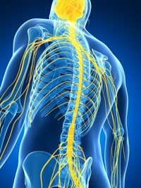
Skin Cells Reprogrammed to Produce Insulin

A cure for type 1 diabetes has alluded researchers. Not because scientists do not know what must be done — but because the tools have not been available to do it. Now scientists at the Gladstone Institutes, harnessing the power of regenerative medicine, have developed a technique in animal models that could replenish the very cells destroyed by the disease. The team’s findings, published online in the journal Cell Stem Cell, are an important step towards freeing an entire generation of patients from the life-long injections that characterize this devastating disease.

Type 1 diabetes, which usually manifests during childhood, is caused by the destruction of ?-cells, a type of cell that normally resides in the pancreas and produces a hormone called insulin. Without insulin, the body’s organs have difficulty absorbing sugars, such as glucose, from the blood. Once a death sentence, the disease can now be managed with regular glucose monitoring and insulin injections. A more permanent solution, however, would be to replace the missing ?-cells. But these cells are hard to come by, so researchers have looked towards stem cell technology as a way to make them.

“The power of regenerative medicine is that it can potentially provide an unlimited source of functional, insulin-producing ?-cells that can then be transplanted into the patient,” said Dr. Ding, who is also a professor at the University of California, San Francisco (UCSF), with which Gladstone is affiliated. “But previous attempts to produce large quantities of healthy ?-cells — and to develop a workable delivery system — have not been entirely successful. So we took a somewhat different approach.”

One of the major challenges to generating large quantities of ?-cells is that these cells have limited regenerative ability; once they mature it’s difficult to make more. So the team decided to go one step backwards in the life cycle of the cell.

The team first collected skin cells, called fibroblasts, from laboratory mice. Then, by treating the fibroblasts with a unique ‘cocktail’ of molecules and reprogramming factors, they transformed the cells into endoderm-like cells. Endoderm cells are a type of cell found in the early embryo, and which eventually mature into the body’s major organs — including the pancreas.

“Using another chemical cocktail, we then transformed these endoderm-like cells into cells that mimicked early pancreas-like cells, which we called PPLC’s,” said Gladstone Postdoctoral Scholar Ke Li, PhD, the paper’s lead author. “Our initial goal was to see whether we could coax these PPLC’s to mature into cells that, like ?-cells, respond to the correct chemical signals and — most importantly — secrete insulin. And our initial experiments, performed in a petri dish, revealed that they did.”

The research team then wanted to see whether the same would occur in live animal models. So they transplanted PPLC’s into mice modified to have hyperglycemia (high glucose levels), a key indicator of diabetes.

“Importantly, just one week post-transplant, the animals’ glucose levels started to decrease gradually approaching normal levels,” continued Dr. Li. “And when we removed the transplanted cells, we saw an immediate glucose spike, revealing a direct link between the transplantation of the PPLC’s and reduced hyperglycemia.”

But it was when the team tested the mice eight weeks post-transplant that they saw more dramatic changes: the PPLC’s had given rise to fully functional, insulin-secreting ?-cells.

“These results not only highlight the power of small molecules in cellular reprogramming, they are proof-of-principle that could one day be used as a personalized therapeutic approach in patients,” explained Dr. Ding.

“I am particularly excited about the prospect of translating these findings to the human system,” said Matthias Hebrok, PhD, one of the study’s authors and director of the UCSF Diabetes Center. “Most immediately, this technology in human cells could significantly advance our understanding of how inherent defects in ?-cells result in diabetes, bringing us notably closer to a much-needed cure.”

So Called Safe Pollution Levels May Not Be

If you’re eating better and exercising regularly, but still aren’t seeing improvements in your health, there might be a reason: pollution. According to a new research report published in the September issue of The FASEB Journal, what you are eating and doing may not be the problem, but what’s in what you are eating could be the culprit. The study described below gives one more very good reason for eating organically raised foods.

“This study adds evidences for rethinking the way of addressing risk assessment especially when considering that the human population is widely exposed to low levels of thousands of chemicals, and that the health impact of realistic mixtures of pollutants will have to be tested as well,” said Brigitte Le Magueresse-Battistoni, a researcher involved in the work from the French National Institute of Health and Medical Research (INSERM). “Indeed, one pollutant could have a different effect when in mixture with other pollutants. Thus, our study may have strong implications in terms of recommendations for food security. Our data also bring new light to the understanding of the impact of environmental food contaminants in the development of metabolic diseases.”

To make this discovery, scientists used two groups of obese mice. Both were fed a high-fat, high-sucrose enriched diet, with one group receiving a cocktail of pollutants added to its diet at a very low dosage. These pollutants were given to the mice throughout — from pre-conception to adulthood. Although the researchers did not observe toxicity or excess of weight gain in the group having received the cocktail of pollutants, they did see a deterioration of glucose tolerance in females, suggesting a defect in insulin signaling. Study results suggest that the mixture of pollutants reduced estrogen activity in the liver through enhancing an enzyme in charge of estrogen elimination. In contrast to females, glucose tolerance was not impacted in males exposed to the cocktail of pollutants. However, males did show some changes in liver related to cholesterol synthesis and transport. This study fuels the concept that pollutants may contribute to the current prevalence of chronic diseases including metabolic diseases and diabetes.

“This report that confirms something we’ve known for a long time: pollution is bad for us,” said Gerald Weissmann, M.D., Editor-in-Chief of The FASEB Journal. “But, what’s equally important, it shows that evaluating food contaminants and pollutants on an individual basis may be too simplistic. We can see that when “safe” levels of contaminants and pollutants act together, they have significant impact on public health.”

Eating the Western Diet May Lead to Premature Death

Data from a new study of British adults suggest that adherence to the Western diet (fried and sweet food, processed and red meat, refined grains, and high-fat dairy products) reduces a person’s likelihood of achieving older ages in good health and with higher functionality. Study results appear in the May issue of The American Journal of Medicine.

“The impact of diet on specific age related diseases has been studied extensively, but few investigations have adopted a more holistic approach to determine the association of diet with overall health at older ages,” says lead investigator Tasnime Akbaraly, PhD, Inserm, Montpellier, France. “We examined whether diet, assessed in midlife, using dietary patterns and adherence to the Alternative Healthy Eating Index (AHEI), is associated with aging phenotypes, identified after a mean 16-year follow-up.”

The AHEI is a validated index of diet quality, originally designed to provide dietary guidelines with the specific intention to combat major chronic conditions such as cardiovascular diseases and diabetes.

Investigators analyzed findings from the British Whitehall II cohort study, which suggest that following the AHEI can double the odds of reversing metabolic syndrome, a condition known to be a strong predictor of heart disease and mortality. The research team sought to identify dietary factors that can not only prevent premature death, but also promote ideal aging.

Researchers followed 3,775 men and 1,575 women from 1985-2009 with a mean age of 51 years from the Whitehall II study. Using a combination of hospital data, results of screenings conducted every five years, and registry data, investigators identified mortality and chronic diseases among participants. The outcomes at follow up stage, classified into 5 categories were:

1. Ideal aging, defined as free of chronic conditions and high performance in physical, mental, and cognitive functioning tests — 4.0%

2. Nonfatal cardiovascular event — 12.7%

3. Cardiovascular death — 2.8%

4. Noncardiovascular death — 7.3%

5. Normal aging — 73.2%

The study determined that participants with low adherence to the AHEI increased their risk of cardiovascular and noncardiovascular death. Those who followed the Western diet consisting of fried and sweet food, processed food and red meat, refined grains, and high-fat dairy products lowered their chances for ideal aging.

“We showed that following specific dietary recommendations such as the one provided by the AHEI may be useful in reducing the risk of unhealthy aging, while avoidance of the ‘Western-type foods’ might actually improve the possibility of achieving older ages free of chronic diseases and remaining highly functional,” notes Dr. Akbaraly. “A better understanding of the distinction between specific health behaviors that offer protection against diseases and those that move individuals towards ideal aging may facilitate improvements in public health prevention packages.”

Exercise Causes Epigenetic Changes to Your Cells

Exercise, even in small doses, changes the expression of our innate DNA. New research from Lund University in Sweden has described for the first time what happens on an epigenetic level in fat cells when we undertake physical activity.

“Our study shows the positive effects of exercise, because the epigenetic pattern of genes that affect fat storage in the body changes,” says Charlotte Ling, Associate Professor at Lund University Diabetes Centre.

The cells of the body contain DNA, which contains genes. We inherit our genes and they cannot be changed. The genes, however, have ‘methyl groups’ attached which affect what is known as ‘gene expression’ — whether the genes are activated or deactivated. The methyl groups can be influenced in various ways, through exercise, diet and lifestyle, in a process known as ‘DNA methylation’. This is epigenetics, a relatively new research field that in recent years has attracted more and more attention.

In the study, the researchers investigated what happened to the methyl groups in the fat cells of 23 slightly overweight, healthy men aged around 35 who had not previously engaged in any physical activity, when they regularly attended spinning and aerobics classes over a six-month period.

“They were supposed to attend three sessions a week, but they went on average 1.8 times,” says Tina R?nn, Associate Researcher at Lund University.

Using technology that analyses 480 000 positions throughout the genome, they could see that epigenetic changes had taken place in 7,000 genes (an individual has 20-25 000 genes). They then went on to look specifically at the methylation in genes linked to type 2 diabetes and obesity.

“We found changes in those genes too, which suggests that altered DNA methylation as a result of physical activity could be one of the mechanisms of how these genes affect the risk of disease,” says Tina R?nn, adding that this has never before been studied in fat cells and that they now have a map of the DNA methylome in fat.

In the laboratory, the researchers were able to confirm the findings in vitro (studying cell cultures in test tubes) by deactivating certain genes and thus reducing their expression. This resulted in changes in fat storage in fat cells.

Adult Stem Cells to Grow New Bone

Preparations are underway for the first known human trial to use stem cells collected from adults to grow new bone.

The cells technology, called VSEL stem cells, or very small embryonic-like stem cells, are derived from adults — not fetuses. This eliminates ethical arguments and potential side effects associated with using actual embryonic stem cells derived from a fetus, say researchers at the University of Michigan School of Dentistry and New York-based NeoStem Inc.

The research partners hypothesize that the VSEL stem cells, which mimic properties of embryonic stem cells, can provide a minimally invasive way to speed painful bone regeneration for dental patients and others with bone trauma.

U-M’s role in the study involves design, patient care and data analysis, while NeoStem provides the cells and patented technology to purify the special stem cells. Study leaders include Russell Taichman, U-M professor of dentistry; Laurie McCauley, professor and newly named dean of the U-M Dental School; and Denis Rodgerson, director of grants and academic liaisons for NeoStem. U-M’s work will take place at the Michigan Center for Oral Health Research and the U-M Health System.

“Within a year, researchers hope to begin recruiting roughly 50 patients who need a tooth extraction and a dental implant,” Taichman said.

Before extracting the tooth, U-M researchers harvest the patient’s cells, and then NeoStem’s VSEL technology is used to purify and isolate those VSEL stem cells from the patient’s other cells.

This allows U-M researchers to implant pure populations of the VSEL stem cells back into test patients. Control patients receive their own cells, not the VSELs. After the new bone grows, researchers remove a small portion of it to analyze, and replace it with an implant.

“We’re taking advantage of the time between extraction and implant to see if these cells will expedite healing time and produce better quality bone,” Taichman said. “They are natural cells that are already in your body, but NeoStem’s technology concentrates them so that we can place a higher quantity of them onto the wound site.”

U-M has applied for initial patent protection to use the VSEL stem cells to grow bone. Robin Smith, chairman and CEO of NeoStem, emphasized the importance of this study for the development of embryonic-like stem cells from the patient’s own body to treat a wide range of diseases.

World’s Oldest Person Reveals Longevity Secret

The world?s oldest living person reached another milestone Sunday when she celebrated her Sweet 116. When asked for her secret to living such a long life she said ?I mind my own business and I don?t eat junk food.?

Besse Cooper has become only the 8th person in the world and the 4th American verified to have turned that age, according to Guinness World Records.

Cooper, whose maiden name was Brown, was born on Aug. 26, 1896, in northern Tennessee the same year as comedian George Burns and writer F. Scott Fitzgerald.

In 1917 during World War I, she moved to Georgia to work as a schoolteacher. She later married Luther Cooper, and they raised four children together. He died in 1963 at age 68, according to The Walton Tribune.

The newspaper reported last year that the supercentenarian is also a grandmother of 12, great-grandmother of 15 and great-great-grandmother of one.

Walton County officials held a ribbon cutting Friday to name a bridge in honor of Cooper, who resides in a nursing home and was unable to attend the ceremony.

?The older she has gotten the more wittier she has gotten,? Cooper?s third child, Sidney, reportedly said, adding that she had a message for the bridge-naming ceremony: ?I?m glad I gave them a reason to name it.?

Although Guinness World Records first called her the world?s oldest living person in January 2011, she lost the title when it was discovered Maria Gomes Valentin, of Brazil, was born 48 days earlier.

Valentin died on June 21, 2011, just shy of her 115th birthday, and Cooper was again crowned the world?s super senior.

Cooper could still surpass another Tennessean, Elizabeth Bolden, who was 116 years and 118 days when she died in 2006.

The person with the longest human lifespan ever recorded was Jeanne Calment, of France, who was born in 1875 and died in 1997 at the age of 122.

Georgia Man’s Foot Regrown After Being Decimated by Flesh-Eating Bacteria

Once they stripped away the infected flesh, the next step was to cut off the remaining foot bone. But Osiris (USA’s leading Repair Stem Cell company) donated its products to see if it could fix the largest problem ever proposed for stem cells anywhere!

Dalton, Georgia May 2012: By the time Dr. Spencer Misner had carved away the dead and diseased flesh from Bobby Rice?s right foot last year, little remained other than bones and tendons.

?I couldn?t believe it. It didn?t look real. It looked like something out of a movie,? recalled Rice.

Today, the ankle has almost completely healed. It looks like Rice had simply scraped it. And Rice?s foot has largely healed, too. Misner credits cutting-edge stem cell treatments for saving Rice?s foot and leg.

Rice, who has diabetes, stepped on a piece of glass last fall and his foot quickly became infected. After trying a home remedy, Rice eventually went to Hamilton Medical Center in Dalton, GA, where doctors found he had a rapidly spreading necrotizing fasciitis, or in layman?s terms, flesh-eating bacteria.

Physicians treated the infection with antibiotics. However, Rice had one toe amputated. Doctors had to strip away much of the flesh from Rice?s foot and a great deal of flesh along his ankle.

?We did what we had to do,? Misner said. ?We got the infection out. We saved his life. But what do you do next? We?d normally say all you can do now is cut of his leg so he can get on with his life.?

But Misner had another idea. He contacted Ed Fickey, a sales representative for Osiris Therapeutics and asked about using the company?s new stem cell technologies to rebuild the foot and ankle.

Stem cells can grow and differentiate into many different types of cells. Stem cell treatments introduce these cells into damaged or diseased organs to repair them.

?The problem is that Bobby is an indigent patient and didn?t have the financial resources. Ed spoke to the company, and they agreed to donate the products for free,? Misner said.

Osiris provided two products called Grafix and Ovation. Fickey said they are made from adult stem cells derived from donated placenta and do not come from embryos.

?They hadn?t had a case of this magnitude before,? Misner said. ?Stem cells are starting to be used pretty extensively. They are used on burn patients to regenerate skin. But this involves muscle, bone, tendon, fat, skin, so many types of tissues.?

Misner said he asked the company for some guidelines.

?They basically said, ?You let is know what the guidelines are because we don?t have a record of anything like this being done before.? I did a literature search, and I couldn?t find anything like this before,? Misner said. ?They did connect me with a doctor in Washington, D.C. who has used stem cells quite a bit. But when I sent him the pictures (of Rice?s foot and ankle), he basically said ?Good luck.? He helped me with some general guidelines, but there was no recipe to follow.?

Misner started the treatments in November 2011, and Fickey recalls just how tricky it was.

?He (Misner) had a syringe, and he was looking for some tissue to push it into. But there was nothing but bone there,? he said. ?Now, there?s a whole fleshy foot.?

Misner has applied the stem cell treatments nine times now, and he describes the foot as more than 90 percent healed. He had to perform the first treatments in an operating room at Hamilton Medical Center because of how complicated they were. But he has been able to do the last few treatments in his office. He expects to have to do at least three more before the foot has completely regenerated.

Fickey said Osiris has been watching ?the Dalton foot? very closely.

?Each time we do an application, I send the latest pictures back, and they have always been very impressed. They wanted to see if there has been muscle growth and the answer has been yes. They wanted to see if there has been vascularization, blood flow, and there has been,? Fickey said. ?The most impressive thing is that Bobby has feeling back. We were here a couple of weeks ago and the nurse tickled his foot and he kind of jumped. We didn?t expect that. Now, we are starting to have some other cases around and we can tell them ?This is what we want to see.??

Rice said he never dreamed he would regain his foot.

?I know what it looked like. I expected he would just have to take it off. To see where it is today is just amazing,? he said.

Hearing Loss Regenerated in Damaged Mammal Ear

Hearing loss is a significant public health problem affecting almost 50 million people in the United States alone. Sensorineural hearing loss is the most common form and is caused by the loss of sensory hair cells in the cochlea. Hair cell loss results from a variety of factors including noise exposure, aging, toxins, infections, and certain antibiotics and anti-cancer drugs. Although hearing aids and cochlear implants can ameliorate the symptoms somewhat, there are no known treatments to restore hearing, because auditory hair cells in mammals, unlike those in birds or fish, do not regenerate once lost. Auditory hair cell replacement holds great promise as a treatment that could restore hearing after loss of hair cells.

In the Jan. 10 issue of Neuron, Massachusetts Eye and Ear and Harvard Medical School researchers demonstrate for the first time that hair cells can be regenerated in an adult mammalian ear by using a drug to stimulate resident cells to become new hair cells, resulting in partial recovery of hearing in mouse ears damaged by noise trauma. This finding holds great potential for future therapeutic application that may someday reverse deafness in humans.

“Hair cells are the primary receptor cells for sound and are responsible for the sense of hearing,” explains senior author, Dr. Albert Edge, of Harvard Medical School and Mass. Eye and Ear. “We show that hair cells can be generated in a damaged cochlea and that hair cell replacement leads to an improvement in hearing.”

In the experiment, the researchers applied a drug to the cochlea of deaf mice. The drug had been selected for its ability to generate hair cells when added to stem cells isolated from the ear. It acted by inhibiting an enzyme called gamma-secretase that activates a number of cellular pathways. The drug applied to the cochlea inhibited a signal generated by a protein called Notch on the surface of cells that surround hair cells. These supporting cells turned into new hair cells upon treatment with the drug. Replacing hair cells improved hearing in the mice, and the improved hearing could be traced to the areas in which supporting cells had become new hair cells.

“The missing hair cells had been replaced by new hair cells after the drug treatment, and analysis of their location allowed us to correlate the improvement in hearing to the areas where the hair cells were replaced,” Dr. Edge said.

This is the first demonstration of hair cell regeneration in an adult mammal. “We’re excited about these results because they are a step forward in the biology of regeneration and prove that mammalian hair cells have the capacity to regenerate,” Dr. Edge said. “With more research, we think that regeneration of hair cells opens the door to potential therapeutic applications in deafness.”

Gene Associated with Active Personality Traits Also Linked to Longevity

A variant of a gene associated with active personality traits in humans seems to also be involved with living a longer life, UC Irvine and other researchers have found.

This derivative of a dopamine-receptor gene — called the DRD4 7R allele — appears in significantly higher rates in people more than 90 years old and is linked to lifespan increases in mouse studies.

Robert Moyzis, professor of biological chemistry at UC Irvine, and Dr. Nora Volkow, a psychiatrist who conducts research at the Brookhaven National Laboratory and also directs the National Institute on Drug Abuse, led a research effort that included data from the UC Irvine-led 90+ Study in Laguna Woods, Calif. Results appear online in The Journal of Neuroscience.

The variant gene is part of the dopamine system, which facilitates the transmission of signals among neurons and plays a major role in the brain network responsible for attention and reward-driven learning. The DRD4 7R allele blunts dopamine signaling, which enhances individuals’ reactivity to their environment.

People who carry this variant gene, Moyzis said, seem to be more motivated to pursue social, intellectual and physical activities. The variant is also linked to attention-deficit/hyperactivity disorder and addictive and risky behaviors.

“While the genetic variant may not directly influence longevity,” Moyzis said, “it is associated with personality traits that have been shown to be important for living a longer, healthier life. It’s been well documented that the more you’re involved with social and physical activities, the more likely you’ll live longer. It could be as simple as that.”

Numerous studies — including a number from the 90+ Study — have confirmed that being active is important for successful aging, and it may deter the advancement of neurodegenerative diseases, such as Alzheimer’s.

Prior molecular evolutionary research led by Moyzis and Chuansheng Chen, UC Irvine professor of psychology & social behavior, indicated that this “longevity allele” was selected for during the nomadic out-of-Africa human exodus more than 30,000 years ago.

In the new study, the UC Irvine team analyzed genetic samples from 310 participants in the 90+ Study. This “oldest-old” population had a 66 percent increase in individuals carrying the variant relative to a control group of 2,902 people between the ages of 7 and 45. The presence of the variant also was strongly correlated with higher levels of physical activity.

Next, Volkow, neuroscientist Panayotis Thanos and their colleagues at the Brookhaven National Laboratory found that mice without the variant had a 7 percent to 9.7 percent decrease in lifespan compared with those possessing the gene, even when raised in an enriched environment.

While it’s evident that the variant can contribute to longevity, Moyzis said further studies must take place to identify any immediate clinical benefits from the research. “However, it is clear that individuals with this gene variant are already more likely to be responding to the well-known medical adage to get more physical activity,” he added.

A Better Treatment for Neck, Back and Joint Pain

Chronic musculoskeletal pain is the number one cause of chronic disability in North America and chronic back pain is the leading cause of disability in Americans under the age of 45. This rampaging epidemic of pain can conceivably be eliminated in 80-90% of sufferers with prolotherapy. This treatment which relies on the body’s own healing process is performed by specially trained medical doctors. It has been shown to be effective for pain associated with: the back, the neck, all joints throughout the body, arthritis, migraines, fibromyalgia, sciatica, herniated discs, tension headaches, sports injuries, fibromyalgia, loose joints, TMJ Syndrome, tendinitis, sciatica and degenerated joints. Many people have been able to avoid spinal surgery or joint replacement with this much safer and often more effective treatment.

Prolotherapy relies on the body’s own healing process to eliminate pain. Most neck, back and other musculoskeletal pain is due to weakness of ligaments and tendons. Since ligaments and tendons are the connective tissue that hold our muscles to bone, and bone to bone, both must be taut and strong.

Back pain results when weak ligaments and tendons cause the spine to become “unstable.” Vertebrae begin to slip, move and rotate from their proper position, causing pressure on the nerves. Limited results in pain alleviation may be achieved with cortisone and other anti-inflammatory agents but these do not address the cause of the pain. Temporary pain suppression is not a cure for the underlying problem: ligament and tendon weakness. Advocates of the technique say Prolotherapy is the long-term solution to chronic pain because it strengthens the ligaments and tendons so they can move the vertebrae back into their proper places.

Prolotherapy involves the injection of an “irritant” solution such as dextrose, salt solution or cod liver oil into the area where the ligaments have either been weakened or damaged through injury, degeneration or aging. The injection is given at the point where the ligament connects to the bone. With this injection, the Prolotherapy doctor causes the body to heal itself through the process of inflammation.

When an irritant is introduced, at the site of injury, the immune system is summoned to the area. The body begins a healing process exactly where the painful area is located. New fibrous tissue is laid, repairing and strengthening the ligaments so that they can pull the vertebrae back where they belong and alleviate pain.

Prolotherapy treatment sessions are generally given every four to six weeks to allow time for the growth of the new connective tissue. Patients usually require four to six treatment sessions for complete recovery, some experience more immediate results.

Standard medical and surgical procedures cannot match Prolotherapys 80-90% effectiveness in eliminating chronic pain, nor can standard medicine match the relative low cost of treatment. Prolotherapy treatments can range from $100-$500, where a typical surgical procedure may cost over 100 times that amount!

There are currently about 300 physicians who practice Prolotherapy in the United States. With the recent rise in popularity, however, this number is expected to multiply greatly within the next few years. A list of doctors in each state can be found at www.getprolo.com