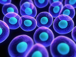
Stem Cells Responsible for Flatworm’s Regenerative Capabilities

After sequencing the genome of flatworms (Macrostomum lignano), researchers from Cold Springs Harbor Laboratory (CSHL) have concluded that it can regenerate parts of its entire body ? with the exception of its brain. According to the scientists, this has potential applications in stem cell research.

“This and other regenerating flatworms have the same kind of pathway operating in stem cells that is responsible for their remarkable regenerative capabilities.” Gregory Hannon, a CSHL professor, said in a news release. “As we started to try to understand the biology of these stem cells, it very quickly became clear that we needed information about the genetic content of these organisms.”

As certain species grow, base cells called stem cells develop into many different cell types. They also act as an internal repair system, dividing and replenishing other cells as needed, similar to how flatworms regenerate body parts that are injured.

Hannon was studying an important pathway in mammalian reproductive tissues when M. lignano caught his eye. When his researchers took a closer look, they found that the flatworm had a very complex genome with repetitive elements, which made it hard to assemble. This required the use of long-read, or high-quality, sequencing technology.

“At the genomic level it has almost no relationship to anything else that’s ever been sequenced. It’s very strange and unique in that sense,” notes Michael Schatz, a CSHL associate professor.

“The worms are just like floating sacks full of stem cells, so they’re very easily accessible,” said Kaja Wasik, the study’s lead author who conducted the work as a Ph.D. student in Hannon’s lab. “From what we looked at, it looks like many of the developmental pathways that are present in humans are also present in the worms, and we can now study whether they potentially could be involved in regeneration.”

M. lignano aids in stem cell researcher because it is small, has simple tissues, is transparent and uses sexual reproduction, the researchers noted. Sequencing the flatworm’s genome is a stepping stone in understanding exactly how its cells are able to regenerate.

Their findings were recently published in the journal Proceedings of the National Academy of Sciences.

Porous Hydrogel May Improve Stem Cell Therapy

Possible stem cell therapies often are limited by low survival of transplanted stem cells and the lack of precise control over their differentiation into the cell types needed to repair or replace injured tissues. A team led by David Mooney, a core faculty member at Harvard?s Wyss Institute, has now developed a strategy that has experimentally improved bone repair by boosting the survival rate of transplanted stem cells and influencing their cell differentiation. The method embeds stem cells into porous, transplantable hydrogels.

In addition to Mooney, the team included Georg Duda, a Wyss associate faculty member and director of the Julius Wolff Institute for Biomechanics and Musculoskeletal Regeneration at Charit? ? Universit?tsmedizin in Berlin, and Wyss Institute founding director Donald Ingber. The team published its findings in today?s issue of Nature Materials. Mooney is also the Robert P. Pinkas Family Professor of Bioengineering at the Harvard John A. Paulson School of Engineering and Applied Sciences.

Stem cell therapies have potential for repairing many tissues and bones, or even for replacing organs. Tissue-specific stem cells can now be generated in the laboratory. However, no matter how well they grow in the lab, stem cells must survive and function properly after transplantation. Getting them to do so has been a major challenge for researchers

Mooney?s team and other researchers have identified specific chemical and physical cues from the stem cell niche (the area in which stem cells survive and thrive with support from other cell types and environmental factors) to promote stem cell survival, multiplication and maturation into tissue. Whereas chemical signals that control stem cell behavior are increasingly understood, much less is known about the mechanical properties of stem cell niches. Stem cells like those present in bone, cartilage, or muscle cultured in laboratories, however, have been found to possess mechanosensitivities, meaning they require a physical substrate with defined elasticity and stiffness to proliferate and mature.

?So far these physical influences had not been efficiently harnessed to propel real-world regeneration processes,? said Nathaniel Huebsch, a graduate student who worked with Mooney and who is the study?s first author. ?Based on our experience with mechanosensitive stem cells, we hypothesized that hydrogels could be leveraged to generate the right chemical and mechanical cues in a first model of bone regeneration.?

Two water-filled hydrogels with very different properties are the key to the Mooney team?s method. A more stable, longer-lasting ?bulk gel? is filled with small bubbles of a second, so-called ?porogen? that degrades at a much faster rate, leaving behind porous cavities.

By coupling the bulk gel with a small ?peptide? derived from the extracellular environment of genuine stem cell niches, and mixing it with a tissue-specific stem cell type as well as the porogen, the team can create a bone-forming artificial niche. While the bulk gel provides just the right amount of elasticity plus a relevant chemical signal to coax stem cells to proliferate and mature, the porogen gradually breaks down, leaving open spaces into which the stem cells expand before they naturally migrate out of the gel structure altogether to form actual mineralized bone tissue.

In small-animal experiments conducted so far, the researchers show that a void-forming hydrogel with the correct chemical and elastic properties provides better bone regeneration than transplanting stem cells alone. Of further interest, the maturing stem cells deployed by the hydrogel also induce nearby native stem cells to contribute to bone repair, further amplifying their regenerative effects.

?This study provides the first demonstration that the physical properties of a biomaterial can not only help deliver stem cells but also tune their behavior in vivo,? said Mooney. ?While so far we have focused on orchestrating bone formation, we believe that our hydrogel concept can be broadly applied to other regenerative processes as well.?

The collaborative, cross-disciplinary work was supported by the Harvard University Materials Research Science and Engineering Center (MRSEC), which is funded by the National Science Foundation (NSF).

?This is an exquisite demonstration of MRSEC programs? high impact,? said Dan Finotello, program director at the NSF. ?MRSECs bring together several researchers of varied experience and complementary expertise who are then able to advance science at a considerably faster rate.?

How Diet Effects Brain Size

New research has shown for the first time that the part of the brain used for learning, memory and mental health is smaller in people with unhealthy diets.

The results of the study by researchers at Deakin University and the Australian National University (ANU) suggest that older Australians with unhealthy diets have smaller hippocampi – the hippocampus is a part of the brain believed to be integral to learning, memory and mental health. It has also shown that older people with healthier diets have larger hippocampi.

Associate Professor Felice Jacka, lead author of the study and researcher with Deakin University’s IMPACT Strategic Research Centre in Geelong, said that as the negative impact of unhealthy foods on the waistline of the population grows, so does the evidence suggesting that our brain health is also affected.

“It is becoming even clearer that diet is critically important to mental as well as physical health throughout life,” Associate Professor Jacka said.

“We’ve known for some time that components of diet, both healthy and unhealthy, have a rapid impact on aspects of the brain that affect hippocampal size and function, but up until now these studies have only been done in rats and mice. This is the first study to show that this also appears to be the case for humans.”

The researchers used magnetic resonance imaging to measure the size of hippocampi (there are two in the brain ? left and right) in Australian adults aged 60-64 years and participating in the PATH study – a large longitudinal study of ageing conducted at the ANU. They also measured the participants’ regular diets and took into account a range of other factors that could affect the hippocampus.

The results of the study, now published in the international journal BMC Medicine, suggest that older adults who eat more unhealthy foods, such as sweet drinks, salty snacks and processed meats, have smaller left hippocampi. It also shows that older adults who eat more nutrient-rich foods, such as vegetables, fruits and fish, have larger left hippocampi. These relationships existed over and above other factors that may explain these associations, such as gender, levels of physical activity, smoking, education or depression itself.

These findings have relevance for both dementia and mental health, Associate Professor Jacka said.

“Mental disorders account for the leading cause of disability worldwide, while rates of dementia are increasing as the population ages,” she said.

“Recent research has established that diet and nutrition are related to the risk for depression, anxiety and dementia, however, until now it was not clear how diet might exert an influence on mental health and cognition.

“This latest study sheds light on at least one of the pathways by which eating an unhealthy diet may influence the risk for dementia, cognitive decline and mental disorders such as depression and anxiety in older people.

“However, it also points to the importance of diet for brain health in other age groups. As the hippocampus is critical to learning and memory throughout life, as well as being a key part of the brain involved in mental health, this study underscores the importance of good nutrition for children, adolescents and adults of all ages.”

Balanced Gut Flora May Improve Age Related Health

Why do some people remain healthy into their 80s and beyond, while others age faster and suffer serious diseases decades earlier? New research led by UCLA life scientists may produce a new way to answer that question and an approach that could help delay declines in health.

Specifically, the study suggests that analyzing intestinal bacteria could be a promising way to predict health outcomes as we age.

The researchers discovered changes within intestinal microbes that precede and predict the death of fruit flies. The findings were published in the open-source journal Cell Reports.

“Age-onset decline is very tightly linked to changes within the community of gut microbes,” said David Walker, a UCLA professor of integrative biology and physiology, and senior author of the research. “With age, the number of bacterial cells increase substantially and the composition of bacterial groups changes.”

The study used fruit flies in part because although their typical life span is just eight weeks, some live to the age equivalent of humans’ 80s and 90s, while others age and die much younger. In addition, scientists have identified all of the fruit fly’s genes and know how to switch individual ones on and off.

In a previous study, the UCLA researchers discovered that five or six days before flies died, their intestinal tracts became more permeable and started leaking.

In the latest research, which analyzed more than 10,000 female flies, the scientists found that they were able to detect bacterial changes in the intestine before the leaking began. As part of the study, some fruit flies were given antibiotics that significantly reduce bacterial levels in the intestine; the study found that the antibiotics prevented the age-related increase in bacteria levels and improved intestinal function during aging.

The biologists also showed that reducing bacterial levels in old flies can significantly prolong their life span.

“When we prevented the changes in the intestinal microbiota that were linked to the flies’ imminent death by feeding them antibiotics, we dramatically extended their lives and improved their health,” Walker said. (Microbiota are the bacteria and other microorganisms that are abundant in humans, other mammals, fruit flies and many other animals.)

Flies with leaky intestines that were given antibiotics lived an average of 20 days after the leaking began — a substantial part of the animal’s life span. On average, flies with leaky intestines that did not receive antibiotics died within a week.

The intestine acts as a barrier to protect our organs and tissue from environmental damage.

“The health of the intestine — in particular the maintenance of the barrier protecting the rest of the body from the contents of the gut — is very important and might break down with aging,” said Rebecca Clark, the study’s lead author. Clark was a UCLA postdoctoral scholar when the research was conducted and is now a lecturer at England’s Durham University.

The biologists collaborated with William Ja, an assistant professor at Florida’s Scripps Research Institute, and Ryuichi Yamada, a postdoctoral research associate in Ja’s laboratory, to produce an additional group of flies that were completely germ-free, with no intestinal microbes. Those flies showed a very dramatic delay in intestinal damage, and they lived for about 80 days, approximately one-and-a-half times as long as the animal’s typical life span.

Scientists have recently begun to connect a wide variety of diseases, including diabetes and Parkinson’s, among many others, to changes in the microbiota, but they do not yet know exactly what healthy microbiota look like.

“One of the big questions in the biology of aging relates to the large variation in how we age and how long we live,” said Walker, who added that scientific interest in intestinal microbes has exploded in the last five years.

When a fruit fly’s intestine begins to leak, its immune response increases substantially and chronically throughout its body. Chronic immune activation is linked with age-related diseases in people as well, Walker said.

Walker said that the study could lead to realistic ways for scientists to intervene in the aging process and delay the onset of Parkinson’s disease, Alzheimer’s disease, cancer, stroke, cardiovascular disease, diabetes and other diseases of aging — although such progress could take many years, he said.

Other co-authors of the research are Matteo Pellegrini, professor of molecular, cell and developmental biology and co-director of UCLA’s Institute for Quantitative and Computational Biosciences, postdoctoral scholars Anna Salazar and Anil Rana, graduate students Jeanette Alcaraz and Marco Morselli, and researcher Sorel Fitz-Gibbon, all of UCLA; and Michael Rera, a former UCLA postdoctoral scholar.

Lack of Sleep Increases Risk of Becoming Sick

Scientists have long associated sufficient sleep with good health. Now they’ve confirmed it.

In 2009, Carnegie Mellon University’s Sheldon Cohen found for the first time that insufficient sleep is associated with a greater likelihood of catching a cold. To do this, Cohen, who has spent years exploring psychological factors contributing to illness, assessed participants self-reported sleep duration and efficiency levels and then exposed them to a common cold virus.

Now, Cohen, the Robert E. Doherty University Professor of Psychology in the Dietrich College of Humanities and Social Sciences, and researchers from UC San Francisco and the University of Pittsburgh Medical Center have confirmed that insufficient sleep is connected to an increased chance of getting sick. Published in the journal Sleep, the researchers used objective sleep measures to show that people who sleep six hours a night or less are more than four times more likely to catch a cold, compared to those who sleep more than seven hours in a night.

Aric Prather, assistant professor of psychiatry at UCSF and lead author of the study, said that the findings add to growing evidence emphasizing how important sleep is for health.

“It goes beyond feeling groggy or irritable,” Prather said. “Not getting enough sleep affects your physical health.”

Cohen’s lab is renowned for using the common cold virus to safely test how various factors affect the body’s ability to fight off disease. Prather approached Cohen about the possibility of investigating sleep and susceptibility to colds using data collected in a recent study in which participants wore sensors to get objective, accurate sleep measures.

“We had worked with Dr. Prather before and were excited about the opportunity to have an expert in the effects of sleep on health take the lead in addressing this important question,” Cohen said.

For the study, 164 adults underwent two months of health screenings, interviews and questionnaires to establish baselines for factors like stress, temperament, and alcohol and cigarette use. The researchers also tracked their sleep patterns for seven days using a watch-like sensor that measured the duration and quality of sleep throughout the night. Then, the participants were sequestered in a hotel, administered the cold virus via nasal drops and monitored for a week, collecting daily mucus samples to see if the virus had taken hold.

They found that subjects who slept less than six hours a night were 4.2 times more likely to catch the cold compared to those who got more than seven hours of sleep, and those who slept less than five hours were 4.5 times more likely.

“Sleep goes beyond all the other factors that were measured,” Prather said. “It didn’t matter how old people were, their stress levels, their race, education or income. It didn’t matter if they were a smoker. With all those things taken into account, statistically sleep still carried the day and was an overwhelmingly strong predictor for susceptibility to the cold virus.”

Prather said the study shows the risks of chronic sleep loss better than typical experiments in which researchers artificially deprive subjects of sleep, because it is based on subjects’ normal sleep behavior.

“This could be a typical week for someone during cold season,” he said.

The study adds another piece of evidence that sleep should be treated as a crucial pillar of public health, along with diet and exercise, the researchers said. But it’s still a challenge to convince people to get more sleep.

“In our busy culture, there’s still a fair amount of pride about not having to sleep and getting a lot of work done,” Prather said. “We need more studies like this to begin to drive home that sleep is a critical piece to our well-being.”

Read about Cohen’s previous study that found insufficient sleep is associated with a greater likelihood of catching a cold at www.cmu.edu/homepage/health/2009/winter/not-sleeping.shtml.

Gene Network Stability Increases Lifespan

Nuts

Age defying species such as the giant red sea urchin, the painted turtle, the naked mole rat and the bowhead whale may begin teaching us how to extend our own lives. All these species stand out because they exhibit negligible senescence, the ability to grow older without suffering functional declines or any age-related increase in mortality. Apparently, these species have something we lack. This something, according to a new study, is gene network stability.

A stabilized gene network promotes longer lifespan. The new data is consistent with Dr. Villeponteau’s model that keeping the optimal gene network at youth from drifting with age generates longer life. Likewise, interventions that nudge gene expression back to that of the youthful gene network reverses aging. That is the goal with all the Life Code products, particularly Stem Cell 100 and Stem Cell 100+.

The new study was undertaken by scientists from the biotech company Gero in collaboration with Robert J. Shmookler Reis, Ph.D., a professor of geriatrics at the University of Arkansas. Dr. Shmookler Reis enjoys the distinction of having extended by a factor of 10 the lifespan of the nematode Caenorhabditis elegans.

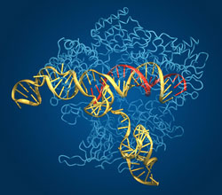
These scientists noticed that in long-lived animals, negligible senescence is accompanied by exceptionally stable gene expression. And stable gene expression, they reasoned, could be attributed to gene network repair systems. To test this idea, the scientists devised a mathematical model of a genetic network. This model not only managed to account for the age-independent mortality exhibited by long-lived animals, it also identified gene network parameters that influence longevity. These parameters include effective gene network connectivity, effective genome size, proteome turnover, and DNA repair rate.

The scientists published their findings August 28 in the journal Scientific Reports, in an article entitled, ?Stability analysis of a model gene network links aging, stress resistance, and negligible senescence.? This article argues that under a very generic set of assumptions, there exist two distinctly different classes of aging dynamics.

?If the repair rates are sufficiently high or the connectivity of the gene network is sufficiently low, then the regulatory network is very stable and mortality is time-independent in a manner similar to that observed in negligibly senescent animals,? wrote the authors. ?Should the repair systems display inadequate efficiency, a dynamic instability emerges, with exponential accumulation of genome-regulation errors, functional declines and a rapid aging process accompanied by an exponential increase in mortality.?

The authors added that the onset of instability depends on the gene-network properties only, irrespective of genotoxic stress levels. Essentially, instability can be viewed as being hard-wired in the genome of the species.

Nonetheless, the authors said that their model implies many possibilities to stabilize a regulatory network and thus extend lifespan. Some of these possibilities, in fact, have already been exploited by nature, accounting for some of the lifespan increases observed to have occurred in evolution. For example, a model parameter that the authors call the coupling rate can be adjusted by increasing or decreasing the degree of gene network connectivity.

A biological embodiment of this parameter is the degree to which the nuclear envelope reduces the effective interactions between the genes and the cellular environment. Once the nuclear envelope became available, organisms that used it enjoyed a dramatic increase in complexity and life expectancy.

Such observations have implications for potential life-extending interventions. For example, experimental reduction of the network connectivity by silencing of kinase cascades involved in regulation of transcription factors leads to a dramatic effect on the lifespan in C. elegans (up to a 10x lifespan extension by a single mutation).

The relation between stresses, stress resistance, and aging, the authors explained, can also account for the way damage to gene regulation from stresses encountered even at a very young age can persist for a very long time and influence lifespan. Such effects, the authors believe, indicate that further research into the relation between gene network stability and aging will make it possible to create entirely new therapies with potentially strong and lasting effect against age-related diseases and aging itself.

Making Safer Stem Cells for Regenerative Medicine

A finding reveals why the transformation process of differentiated cells into stem cells results in significant damage to the DNA. Researchers have managed to rectify this damage using a simple modification to the culture medium, which produces potentially safer stem cells for use in regenerative medicine.

Damaged tissue, such as pancreas, heart, and neuronal tissue, which is regenerated to treat cardiovascular diseases, diabetes, or neurodegenerative diseases. This is one of the ambitious scenarios to which regenerative medicine aspires and that is being announced as one of the great promises of twenty-first century biomedicine for the treatment of a long list of diseases affecting people today. The focal point is the use of stem cells, which are capable of producing different types of cells or tissue.

2006 marked a turning point in this field, when the Japanese scientist, Shinya Yamanaka, managed to generate pluripotent stem cells in the lab for the first time. These are capable of becoming any type of cell, whether insulin-producing beta cells (pancreas) or cardiomyocytes (heart), and are known as iPS cells. This cell reprogramming technique eliminated one of the great ethical dilemmas of the time: until then, pluripotent stem cells could only be obtained from embryos which, in order to achieve this, had to be destroyed.

However, as ?scar Fern?ndez-Capetillo, head of the Genomic Instability Group at the Spanish National Cancer Research Centre (CNIO), says: “the drawback of this new technology is that Yamanaka’s method damaged the stem cell genome, leading to certain safety concerns regarding these cells.” While the fact that the method damaged the DNA of iPS cells was known, the reasons were not.

According to an article published this week in Nature Communications, the team headed by Fern?ndez-Capetillo states that the damage to the genome of iPS cells lies in a very specific kind of stress that the cells are subjected to during cell reprogramming: replication stress, which occurs when the cells increase the pace of division. In addition, and based on these findings, the authors of the paper have managed to develop strategies to reduce this type of stress, resulting in pluripotent stem cells with less damage to their genome.

The results represent a significant step forward regarding the possible use of iPS cells, because after almost a decade since they were developed, there is now a more efficient way of obtaining them, with less damage to the DNA, making them potentially safer.

The CNIO Telomeres and Telomerase groups, headed by Mar?a Blasco, and the Tumoral Suppression Group, headed by Manuel Serrano have also participated in this study, together with groups from the Pasteur Institute in Paris, Toronto University and the Pompeu Fabra University in Barcelona.

Stem Cells With More Stable Genomes

The nature of the damage to the DNA observed in iPS cells has been intensely discussed for some years, due to the fact that it is linked to the rearrangement of large fragments of chromosomes which could lead to potentially dangerous mutations if used clinically.

In a paper published in Nature in 2009, the team led by Mar?a Blasco, with the collaboration of Fern?ndez-Capetillo’s group, described how the damage to the DNA had important consequences in cell reprogramming by limiting the process and making it less efficient.

Now the team headed by Fern?ndez-Capetillo has not only identified the origin of the damage, replication stress, but has managed to reduce it significantly; potentially improving the safety of induced stem cells for use in biomedicine.

To reduce damage to stem cells and thus achieve more stable genomes, the scientists have used a dual approach: genetics, increasing the production of the Chk1 protein, which repairs DNA damage due to replication stress; and chemical, based on supplementing the medium in which the cells are fed with nucleoside, the source compounds of the bricks that build DNA.

“Based on previous research performed by the group, we knew that an additional input of nucleoside reduces replication stress, probably by facilitating the successful replication of DNA as it increases the rate of cell division during the reprogramming process,” explains Sergio Ruiz, whose signature appear in first place on the paper.

The simplicity of this nucleoside-based strategy means that it can be implemented easily by laboratories around the world working with iPS cells, and thus contribute significantly to the field of regenerative biology, one of the greatest aspirations of biomedicine this century.

Journal Reference:

1.Sergio Ruiz, Andres J. Lopez-Contreras, Mathieu Gabut, Rosa M. Marion, Paula Gutierrez-Martinez, Sabela Bua, Oscar Ramirez, I?igo Olalde, Sara Rodrigo-Perez, Han Li, Tomas Marques-Bonet, Manuel Serrano, Maria A. Blasco, Nizar N. Batada, Oscar Fernandez-Capetillo. Limiting replication stress during somatic cell reprogramming reduces genomic instability in induced pluripotent stem cells. Nature Communications, 2015; 6: 8036 DOI: 10.1038/ncomms9036

Exercise Improves Brain Health

One day soon, doctors may determine how physically active you are simply by imaging your brain. Physically fit people tend to have larger brain volumes and more intact white matter than their less-fit peers. Now a new study reveals that older adults who regularly engage in moderate to vigorous physical activity have more variable brain activity at rest than those who don’t. This variability is associated with better cognitive performance, researchers say.

The new findings are reported in the journal PLOS ONE.

“We looked at 100 adults between the ages of 60 and 80, and we used accelerometers to objectively measure their physical activity over a week,” said University of Illinois postdoctoral researcher Agnieszka Burzynska, who led the study with Beckman Institute for Advanced Science and Technology director Art Kramer.

The researchers also used functional MRI to observe how blood oxygen levels changed in the brain over time, reflecting each participant’s brain activity at rest. And they evaluated the microscopic integrity of each person’s white-matter fibers, which carry nerve impulses and interconnect the brain.

“We found that spontaneous brain activity showed more moment-to-moment fluctuations in the more-active adults,” said Burzynska, who now is a professor at Colorado State University. “In a previous study, we showed that in some of the same regions of the brain, those people who have higher brain variability also performed better on complex cognitive tasks, especially on intelligence tasks and memory.”

The researchers also found that, on average, older adults who were more active had better white-matter structure than their less-active peers.

“Our study, when viewed in the context of previous studies that have examined behavioral variability in cognitive tasks, suggests that more-fit older adults are more flexible, both cognitively and in terms of brain function, than their less-fit peers,” Kramer said.

The new research highlights yet another way to assess brain health in aging, Burzynska said.

“We want to know how the brain relates to the body, and how physical health influences mental and brain health in aging,” she said. “Here, instead of a structural measure, we are taking a functional measure of brain health. And we are finding that tracking changes in blood-oxygenation levels over time is useful for predicting cognitive functioning and physical health in aging.”

Exercise Improves Brain Function In Older Adults

One day soon, doctors may determine how physically active you are simply by imaging your brain. Physically fit people tend to have larger brain volumes and more intact white matter than their less-fit peers. Now a new study reveals that older adults who regularly engage in moderate to vigorous physical activity have more variable brain activity at rest than those who don’t. This variability is associated with better cognitive performance, researchers say.

The new findings are reported in the journal PLOS ONE.

“We looked at 100 adults between the ages of 60 and 80, and we used accelerometers to objectively measure their physical activity over a week,” said University of Illinois postdoctoral researcher Agnieszka Burzynska, who led the study with Beckman Institute for Advanced Science and Technology director Art Kramer.

The researchers also used functional MRI to observe how blood oxygen levels changed in the brain over time, reflecting each participant’s brain activity at rest. And they evaluated the microscopic integrity of each person’s white-matter fibers, which carry nerve impulses and interconnect the brain.

“We found that spontaneous brain activity showed more moment-to-moment fluctuations in the more-active adults,” said Burzynska, who now is a professor at Colorado State University. “In a previous study, we showed that in some of the same regions of the brain, those people who have higher brain variability also performed better on complex cognitive tasks, especially on intelligence tasks and memory.”

The researchers also found that, on average, older adults who were more active had better white-matter structure than their less-active peers.

“Our study, when viewed in the context of previous studies that have examined behavioral variability in cognitive tasks, suggests that more-fit older adults are more flexible, both cognitively and in terms of brain function, than their less-fit peers,” Kramer said.

The new research highlights yet another way to assess brain health in aging, Burzynska said.

“We want to know how the brain relates to the body, and how physical health influences mental and brain health in aging,” she said. “Here, instead of a structural measure, we are taking a functional measure of brain health. And we are finding that tracking changes in blood-oxygenation levels over time is useful for predicting cognitive functioning and physical health in aging.”

Sleep Position Affects Brain Health

side sleeping

Sleeping in the lateral, or side position, as compared to sleeping on one’s back or stomach, may more effectively remove brain waste and prove to be an important practice to help reduce the chances of developing Alzheimer’s, Parkinson’s and other neurological diseases, according to researchers at Stony Brook University.

By using dynamic contrast magnetic resonance imaging (MRI) to image the brain’s glymphatic pathway, a complex system that clears wastes and other harmful chemical solutes from the brain, Stony Brook University researchers Hedok Lee, PhD, Helene Benveniste, MD, PhD, and colleagues, discovered that a lateral sleeping position is the best position to most efficiently remove waste from the brain. In humans and many animals the lateral sleeping position is the most common one. The buildup of brain waste chemicals may contribute to the development of Alzheimer’s disease and other neurological conditions. Their finding is published in the Journal of Neuroscience.

Dr. Benveniste, Principal Investigator and a Professor in the Departments of Anesthesiology and Radiology at Stony Brook University School of Medicine, has used dynamic contrast MRI for several years to examine the glymphatic pathway in rodent models. The method enables researchers to identify and define the glymphatic pathway, where cerebrospinal fluid (CSF) filters through the brain and exchanges with interstitial fluid (ISF) to clear waste, similar to the way the body’s lymphatic system clears waste from organs. It is during sleep that the glymphatic pathway is most efficient. Brain waste includes amyloid ? (amyloid) and tau proteins, chemicals that negatively affect brain processes if they build up.

In the paper, “The Effect of Body Posture on Brain Glymphatic Transport,” Dr. Benveniste and colleagues used a dynamic contrast MRI method along with kinetic modeling to quantify the CSF-ISF exchange rates in anesthetized rodents’ brains in three positions ? lateral (side), prone (down), and supine (up).

“The analysis showed us consistently that glymphatic transport was most efficient in the lateral position when compared to the supine or prone positions,” said Dr. Benveniste. “Because of this finding, we propose that the body posture and sleep quality should be considered when standardizing future diagnostic imaging procedures to assess CSF-ISF transport in humans and therefore the assessment of the clearance of damaging brain proteins that may contribute to or cause brain diseases.”

Dr. Benveniste and first-author Dr. Hedok Lee, Assistant Professor in the Departments of Anesthesiology and Radiology at Stony Brook developed the safe posture positions for the experiments. Their colleagues at the University of Rochester, including Lulu Xie, Rashid Deane and Maiken Nedergaard, PhD, used fluorescence microscopy and radioactive tracers to validate the MRI data and to assess the influence of body posture on the clearance of amyloid from the brains.

“It is interesting that the lateral sleep position is already the most popular in human and most animals ? even in the wild ? and it appears that we have adapted the lateral sleep position to most efficiently clear our brain of the metabolic waste products that built up while we are awake,” says Dr. Nedergaard. “The study therefore adds further support to the concept that sleep subserves a distinct biological function of sleep and that is to ‘clean up’ the mess that accumulates while we are awake. Many types of dementia are linked to sleep disturbances, including difficulties in falling asleep. It is increasing acknowledged that these sleep disturbances may accelerate memory loss in Alzheimer’s disease. Our finding brings new insight into this topic by showing it is also important what position you sleep in,” she explained.

Dr. Benveniste cautioned that while the research team speculates that the human glymphatic pathway will clear brain waste most efficiency when sleeping in the lateral position as compared to other positions, testing with MRI or other imaging methods in humans are a necessary first step.

Reference: “The Effect of Body Posture on Brain Glymphatic Transport” The Journal of Neuroscience, 5 August 2015, 35(31): 11034-11044; DOI: 10.1523/JNEUROSCI.1625-15.2015