
How Many Steps Per Day For a Longer Life?
 For years 10,000 steps per day has been the target for many people! But there is actually nothing magical about it! The original basis for establishing that number was never based on scientific basis. In fact it was part of a marketing campaign launched in Japan in1965 to help promote a pedometer. And it is a big number that many people find hard to reach!
Dr. I-Min Lee at Brigham and Women’s Hospital along with a professor at Harvard Medical School and a researcher on physical activity, set out to discover the basis for 10,000 steps and also study its validity. Their study published in JAMA Internal Medicine set out to answer 2 questions about mortality…how many steps per day are associated with lowering the mortality rate and does stepping up the intensity level make a difference with the same number of steps?
A study was designed that included about 17,000 older women with the average age of 72. This group tends to be less active and health issues occur and become more important as people age. Between 2011 and 2015, the participants wore tracking devices during waking hours to track their steps as they went about their daily activities.
Key findings showed that sedentary women averaged 2,700 step per day. The women who averaged 4,400 steps per day had a 41% reduction in mortality. Mortality rates continued to improve progressively before leveling off at bout 7,500 steps per day. About nine fewer deaths occurred per 1,000 person years among the most active group compared to the least active group.
The research shows that if mortality is a person’s major concern, then the study suggests that you can reap benefits from 7,500 steps per day. That is 25% fewer steps than the common goal of 10,000 steps. The study indicates that even light walking can result in benefits for older women.
The study was designed to look at just two factors. Since it was about mortality, it did not relate steps per day to anything related to cognitive functions, quality of life, or physical conditions. It does not tell us how many steps are needed in order to maximize those things. And in answer to the researchers second question, they did not find that the intensity of the steps mattered.
To view the original scientific study click below:
Association of Step Volume and Intensity With All-Cause Mortality in Older Women
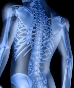 Researchers at Baylor College of Medicine have revealed a new mechanism that will contribute to adult bone maintenance and repair. This new development opens up the possibility for developing new therapeutic strategies for the improvement of bone healing.
 New research has provided the first evidence that along with pollution, smoking, obesity, and diesel exhaust, oxidative stress acts directly on telomeres to speed up cellular aging. The research has shown how stress can cause our biochemical body clock built into our chromosomes to tick faster.
 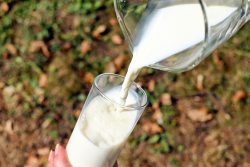 A study conducted at Brigham Young University by exercise scientists has found that people who consume low fat milk experience several years less biological aging when compared to those who drink high fat milk, both 2% and whole milk. This supports existing dietary guidelines which do not recommend high fat milk as part of a healthy diet.
  Scientists have identified pathways that could enable humans to live for well over 100 healthy years according to one of the scientists who participated in the research. The synergistic cellular pathways for longevity that increase the lifespan five fold were discovered in C. elegans which are a nematode worm used as models in aging research. This translates to extending human lifespan by about 50%.
 New research has revealed how a high level of exercise can slow one type of aging. This is the kind of aging that occurs within our cells. We just have to be willing to put in some sweat equity!
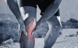 Due to the buildup of scar tissue from a variety of tendon injuries such as jumper’s knee and rotator cuffs, secondary tendon ruptures along with painful and challenging recoveries can occur. New research has shown that the existence of stem cells in tendons can potentially be harnessed to not only improve the healing of tendons but also even avoid surgery.
 A new study by researchers at the University of Sydney’s Faculty of Engineering and IT have found that compounds contained in elderberries can directly inhibit the flu virus’s entry and also replication in human cells. These compounds can additionally help strengthen the immune response to this virus. While modern scientists have struggled to explain the medicinal benefits of herbal products and folk medicine, these methods have been used for millennia to help combat a variety of ailments.
 Typically for many people a New Year is the perfect time to adopt new habits both in the gym and at the grocery store. People can be eager to try out new diets, but does scientific evidence actually support claims made for these diets?
 Researchers from the University of Illinois have conducted a new study on rats that suggests caffeine may offset some of the health risks associated with diets high in sugar and fat. What they found was that rats who consumed caffeine which was extracted from mate tea gained 16% less weight and accumulated 22% less body fat compared to rats who consumed decaffeinated mate tea.