
Why Average Body Temperature has Dropped in Healthy Adults
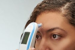 Almost two centuries ago the current “normal” body temperature in humans was established at 98.6 degrees Fahrenheit and has been used as the measure for assessing fevers. However, over time lowering of body temperature has been widely substantiated in healthy adults.
A study conducted in 2017 among 35,000 adults in the U. K. found average body temperatures to be lower at 97.9F and a study in 2019 in the U. S. found normal body temperatures at about 97.5F.
Interestingly, recent studies in both the U. S. and the U. K. have found a decrease similar in the Tsimane which is an indigenous population of forager horticulturists located in the Bolivian Amazon. A team that has been observing this population have observed a rapid decline in body temperature of 0.09 degrees Fahrenheit such that today this population’s body temperatures are about 97.7F.
In less than twenty years researchers are seeing the same levels of decline as that which has been observed in the U. S. over about two centuries. The analysis is based on a large sample of 18,000 observations of about 5,500 adults with adjustments for factors that can affect body temperature such as body mass and ambient temperature.
It is clear that something about human physiology has changed. The researchers leading hypothesis is that we have recorded fewer infections due to clean water, improved hygiene, medical treatment and vaccinations. In the recent study, they were able to test these ideas directly. They have information on clinical diagnosis and also biomarkers of inflammation and infection at the time each participant had been seen.
Although some infections were associated with high body temperatures, adjusting for those did not account for the steep decline in temperatures over time. The same kind of thermometer was used for most of the study so the changes were not due to changes in instruments.
No matter how they did the analysis, the declines were still there. Even when they restricted analysis to under 10% of adults who were diagnosed as completely healthy, they still observed the same decline in body temperatures over time.
A key question became then is why are body temperatures declining in both Americans and the Tsimane population. Data available from the team’s long-term research in Bolivia addressed some of the possibilities. Declines could be due to an increase of modern health care and less incidence of lingering mild infections compared to the past. However, while health has improved over the past two decades, infections are still widespread in rural areas of Bolivia. This then suggests that reduced infection alone isn’t why the decline in body temperatures.
It may be that people are in general in better condition which means their bodies could be working less to fight infection. Greater access to antibiotics and other medical treatments means that the duration of infections is shorter now than was in the past. The team did find that respiratory infection in the early period of the study led to having a high body temperature than having the same infection more recently.
Another possible explanation is that a person’s body doesn’t have to work as hard to maintain the internal temperature due to air conditioning in the warmer temperatures and heating in the colder temperatures. Although Tsimane body temperatures will change with time of year and patterns of the weather, these people do not use any advanced technology for helping to regulate their body temperature. They do however, have more access to blankets and clothes.
The research team was initially surprised to find no single magic bullet that would explain the decline in body temperature. They believe it is likely due to a combination of factors which all point to improved conditions.
Temperature as a vital sign is an indicator of what is occurring physiologically inside the body. One thing that has been known for a while is that there is not universal body temperature for everyone at all times. Despite the fixation on 98.6F, most clinicians know that normal temperatures have a range and throughout the day, temperatures can vary by as much as 1 degree Fahrenheit from lowest in the morning to its highest in the afternoon. It will also vary following physical activity, across the menstrual cycle, and tends to decrease with age.
Through linking improvements in the broader epidemiological and socioeconomic landscape to body temperature changes, the study does suggest that information in regards to body temperature may provide clues to a population’s overall health along with other normal expectations such as life expectancy. Since body temperature is easy to measure, it can easily be added to routine large-scale surveys that monitor the health of populations.
To view the original scientific study click below
Rapidly declining body temperature in a tropical human population.
 A new study has shown that people who are cheerful and feel enthusiastic or what is known as the “positive effect”, are not likely to experience decline in memory with aging. This study adds to a increasing body of research that shows the role a positive outlook has on aging.
 New research has identified a mechanism that appears to be able to prevent hair loss. A group of researchers in Helsinki and Cologne Germany have demonstrated that a protein known as Rictor holds a important role in the process.
  A research team at the Icahn School of Medicine at Mt. Sinai have identified certain sub-populations of brain cells located in the prefrontal cortex that are needed for normal sociability in adults and are also profoundly vulnerable to social isolation in juvenile mice. The prefrontal cortex in the brain is a key part of the brain that regulates social behavior. The study conducted on mice shows long lasting effects and also directs the way to potential treatments.
 Nano-particles can be useful and even valuable in a variety of products, however according to a recent study they can also damage our cells. Researchers are now concerned about the effects of lifelong exposures to this human organism.
 The death of neurons whether in the eye or in the brain can result in a variety of human neurodegenerative diseases that range from blindness to Parkinson’s disease. Treatments currently available for these type of disorders only slow the progression of the illness. This is because once a neuron dies it cannot be replaced.
 Research from the Centre for Nutrition, Exercise and Metabolism at the University of Bath (UK) looked at the combined effects of caffeine and disrupted sleep on our metabolism and found surprising results. They found that a strong, black cup of coffee to wake you up following a bad night’s sleep might impair control of blood sugar levels.
 A new study conducted on mice has suggested that a diet that is high in the sugar fructose exacerbates inflammatory bowel disease (IBD). It appears that changes in gut bacteria mediate the effect.
 According to the CDC at least 5 million people living in the U.S. live with Alzheimer’s Disease and related dementias. As the population ages, experts believe this number will increase quite significantly. A new study of older adults has shown that how increasing physical activity and changing diet can reduce the risk of dementia related diseases even if the person already has a cognitive decline diagnosis.
 Researchers at the Karolinska Institutet have recently revealed new knowledge on the cellular makeup and growth of teeth that can expedite new developments in regenerative dentistry and treatments for tooth sensitivity. Regenerative dentistry is a biological therapy for damaged teeth.