Exercise For Long Telomeres, Too Much Sitting For Short Telomeres

Telomeres

A recent study showed that “cells age faster with a sedentary lifestyle. Chronological age doesn’t always match biological age,” according to Aladdin Shadyab, PhD, lead author of the study with the Department of Family Medicine and Public Health at UC San Diego School of Medicine.

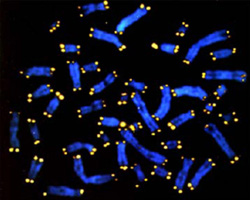
Telomeres are tiny caps found on the ends of DNA strands, like the plastic tips of shoelaces, that protect chromosomes from deterioration. As a cell ages, its telomeres naturally shorten and fray, but health and lifestyle factors, such as obesity and smoking, may accelerate that process.

The study, publishing online January 18 in the American Journal of Epidemiology, found elderly women with less than 40 minutes of moderate-to-vigorous physical activity per day and who remain sedentary for more than 10 hours per day have shorter telomeres.

The researchers also found that elderly women who sit for more than 10 hours a day with low physical activity have cells that are biologically older by eight years compared to women who are less sedentary.

Shadyab and his research team believe they are the first to objectively measure how the combination of sedentary time and exercise can impact the aging biomarker.

Nearly 1,500 women, ages 64 to 95, participated in the study. The women are part of the larger Women’s Health Initiative (WHI), a national, longitudinal study investigating the determinants of chronic diseases in postmenopausal women. The participants completed questionnaires and wore an accelerometer on their right hip for seven consecutive days during waking and sleeping hours to track their movements.

“We found that women who sat longer did not have shorter telomere length if they exercised for at least 30 minutes a day, the national recommended guideline,” said Shadyab. “Discussions about the benefits of exercise should start when we are young, and physical activity should continue to be part of our daily lives as we get older, even at 80 years old.”

Shadyab said future studies will examine how exercise relates to telomere length in younger populations and in men.

Reference: Aladdin H. Shadyab et al. Associations of Accelerometer-Measured and Self-Reported Sedentary Time With Leukocyte Telomere Length in Older Women. American Journal of Epidemiology, January 2017 DOI: 10.1093/aje/kww196

Abstract: Few studies have assessed the association of sedentary time with leukocyte telomere length (LTL). In a cross-sectional study conducted in 2012?2013, we examined associations of accelerometer-measured and self-reported sedentary time with LTL in a sample of 1,481 older white and African-American women from the Women’s Health Initiative and determined whether associations varied by level of moderate- to vigorous-intensity physical activity (MVPA). The association between sedentary time and LTL was evaluated using multiple linear regression models. Women were aged 79.2 (standard deviation, 6.7) years, on average. Self-reported sedentary time was not associated with LTL. In a model adjusting for demographic characteristics, lifestyle behaviors, and health-related factors, among women at or below the median level of accelerometer-measured MVPA, those in the highest quartile of accelerometer-measured sedentary time had significantly shorter LTL than those in the lowest quartile, with an average difference of 170 base pairs (95% confidence interval: 4, 340). Accelerometer-measured sedentary time was not associated with LTL in women above the median level of MVPA. Findings suggest that, on the basis of accelerometer measurements, higher sedentary time may be associated with shorter LTL among less physically active women.

Eat Hot Red Chili Peppers to Increase Life Span

Red Chili Peppers

Like spicy food? If so, you might live longer, say researchers at the Larner College of Medicine at the University of Vermont, who found that consumption of hot red chili peppers is associated with a 13 percent reduction in total mortality in a large prospective study.

For centuries, peppers and spices have been thought to improve health and longevity, but only one other scientific study conducted in China and published in 2015 has previously examined chili pepper consumption and its association with mortality. This new study corroborates the earlier study’s findings.

Using National Health and Nutritional Examination Survey (NHANES) III data collected from more than 16,000 Americans who were followed for up to 23 years, medical student Mustafa Chopan ’17 and Professor of Medicine Benjamin Littenberg, M.D., examined the baseline characteristics of the participants according to hot red chili pepper consumption. They found that consumers of hot red chili peppers tended to be “younger, male, white, Mexican-American, married, and to smoke cigarettes, drink alcohol, and consume more vegetables and meats,” in comparison to participants who did not consume red chili peppers. They examined data from a median follow-up of 18.9 years and observed the number of deaths and then analyzed specific causes of death.

“Although the mechanism by which peppers could delay mortality is far from certain, Transient Receptor Potential (TRP) channels, which are primary receptors for pungent agents such as capsaicin (the principal component in chili peppers), may in part be responsible for the observed relationship,” say the study authors.

There are some possible explanations for red chili peppers’ health benefits, state Chopan and Littenberg in the study. Among them are the fact that capsaicin is believed to play a role in cellular and molecular mechanisms that prevent obesity and modulate coronary blood flow, and also possesses antimicrobial properties that “may indirectly affect the host by altering the gut microbiota.”

“Because our study adds to the generalizability of previous findings, chili pepper or even spicy food consumption may become a dietary recommendation and/or fuel further research in the form of clinical trials,” says Chopan.

Reference: Mustafa Chopan, Benjamin Littenberg. The Association of Hot Red Chili Pepper Consumption and Mortality: A Large Population-Based Cohort Study. PLOS ONE, 2017; 12 (1): e0169876 DOI: 10.1371/journal.pone.0169876

Abstract: “The evidence base for the health effects of spice consumption is insufficient, with only one large population-based study and no reports from Europe or North America. Our objective was to analyze the association between consumption of hot red chili peppers and mortality, using a population-based prospective cohort from the National Health and Nutritional Examination Survey (NHANES) III, a representative sample of US noninstitutionalized adults, in which participants were surveyed from 1988 to 1994. The frequency of hot red chili pepper consumption was measured in 16,179 participants at least 18 years of age. Total and cause-specific mortality were the main outcome measures. During 273,877 person-years of follow-up (median 18.9 years), a total of 4,946 deaths were observed. Total mortality for participants who consumed hot red chili peppers was 21.6% compared to 33.6% for those who did not (absolute risk reduction of 12%; relative risk of 0.64). Adjusted for demographic, lifestyle, and clinical characteristics, the hazard ratio was 0.87 (P = 0.01; 95% Confidence Interval 0.77, 0.97). Consumption of hot red chili peppers was associated with a 13% reduction in the instantaneous hazard of death. Similar, but statistically nonsignificant trends were seen for deaths from vascular disease, but not from other causes. In this large population-based prospective study, the consumption of hot red chili pepper was associated with reduced mortality. Hot red chili peppers may be a beneficial component of the diet.”

More Physical Activity For A Better Brain

Walking my way to 100

Like everything else in the body, the white-matter fibers that allow communication between brain regions also decline with age. In a new study, researchers found a strong association between the structural integrity of these white-matter tracts and an older person’s level of daily activity not just the degree to which the person engaged in moderate or vigorous exercise, but also whether he or she was sedentary the rest of the time.

The study, reported in the journal PLOS ONE, tracked physical activity in 88 healthy but “low-fit” participants aged 60 to 78. The participants agreed to wear accelerometers during most of their waking hours over the course of a week, and also submitted to brain imaging.

“To our knowledge, this is the first study of its kind that uses an objective measure of physical activity along with multiple measures of brain structure,” said University of Illinois postdoctoral researcher Agnieszka Burzynska, who conducted the research with U. of I. Beckman Institute director Arthur Kramer and kinesiology and community health professor Edward McAuley.

Most studies ask subjects to describe how much physical activity they get, which is subjective and imprecise, Burzynska said. The accelerometer continuously tracks a person’s movement, “so it’s not what they say they do or what they think they do, but we have measured what they are actually doing,” she said.

The researchers assumed that participants’ activity levels over a week accurately reflected their overall engagement, or lack of engagement, in physical activity.

The study also relied on two types of brain imaging. The first, diffusion tensor imaging, offers insight into the structural integrity of a tissue by revealing how water is diffused in the tissue. The second method looks for age-related changes in white matter, called lesions. Roughly 95 percent of adults aged 65 and older have such lesions, Burzynska said. While they are a normal part of aging, their early onset or rapid accumulation may spell trouble, she said.

The team found that the brains of older adults who regularly engaged in moderate-to-vigorous exercise generally “showed less of the white-matter lesions,” Burzynska said.

The association between physical activity and white-matter structural integrity was region-specific, the researchers reported. Older adults who engaged more often in light physical activity had greater structural integrity in the white-matter tracts of the temporal lobes, which lie behind the ears and play a key role in memory, language, and the processing of visual and auditory information.

In contrast, those who spent more time sitting had lower structural integrity in the white-matter tracts connecting the hippocampus, “a structure crucial for learning and memory,” Burzynska said.

“This relationship between the integrity of tracts connecting the hippocampus and sedentariness is significant even when we control for age, gender and aerobic fitness,” she said. “It suggests that the physiological effect of sitting too much, even if you still exercise at the end of the day for half an hour, will have a detrimental effect on your brain.”

The findings suggest that engaging in physical activity and avoiding a sedentary lifestyle are both important for brain health in older age, Burzynska said.

“We hope that this will encourage people to take better care of their brains by being more active,” she said.

Reference: Agnieszka Zofia Burzynska ,Laura Chaddock-Heyman,Michelle W. Voss,Chelsea N. Wong,Neha P. Gothe,Erin A. Olson,Anya Knecht,Andrew Lewis,Jim M. Monti,Gillian E. Cooke,Thomas R. Wojcicki,Jason Fanning,Hyondo David Chung,Elisabeth Awick,Edward McAuley,Arthur F. Kramer. Physical Activity and Cardiorespiratory Fitness Are Beneficial for White Matter in Low-Fit Older Adults. PLoS ONE 9(9): e107413. doi:10.1371/journal.pone.0107413

Stem Cells Regrow Damaged Teeth

healthy-teeth

The stem cells in our teeth can be energized to fill in chips, cracks, and cavities, researchers say, and the findings could one day possibly make dental cement obsolete.

The work has been conducted just in mice so far, but the research, published Monday in the journal Scientific Reports, highlights a way to motivate stem cells to repair tooth defects at a scale they normally can?t, with a drug that already has some safety testing behind it. It also demonstrates the potential of a type of stem cell therapy in which the cells are stimulated in place, rather than taken out, manipulated, and put back in.

?We?re mobilizing stem cells in the body and it works,? said Paul Sharpe, a researcher at King?s College London and an author of the new paper. ?If it works for teeth, chances are it could work for other organs.?

Experts not involved with the work noted that while it is in early stages, the simplicity of the approach should ease its path into the next phases of research that show whether it might produce the same results in people.

?These important steps close down the translational gap and bring this discovery a step closer to future clinical applications,? Dr. Vanessa Chrepa, a researcher at the University of Washington, wrote in an email. ?This work will hopefully set the stage for clinical studies in the near future.?

When teeth lose some of their dentin ? the bony tissue beneath the enamel that makes up the bulk of the tooth ? the stem cells tucked deep inside mount a recovery effort and manufacture new dentin (which is also spelled dentine). The problem, Sharpe said, is that the natural repair mechanism can only regrow small amounts of dentin and can?t make up all that is lost when a tooth suffers a serious injury, contracts a major infection, or takes on the sharp end of a dentist?s drill.

Because of the limits of the teeth?s ability to repair themselves, dentists have to fill or seal teeth to prevent further infection and degradation. But dental cement also prevents the tooth from ever returning to its natural, pearly white self.

Sharpe and his team have been trying to understand how the natural repair mechanism works in hopes of converting that understanding into a way to super-power it. As part of their research, they discovered that a group of molecules called glycogen synthase kinase inhibitors (or GSK-3 inhibitors) boosts the stem cells? ability to stimulate production of dentin beyond what normally occurs.

For the new study, the researchers drilled tiny holes into mice?s molars to expose the tooth?s pulp, where the stem cells live. They then inserted collagen sponges that had been soaked in one of three types of GSK-3 inhibitors and covered the tooth.

After six weeks, the researchers removed the teeth and found that the sponges had dissolved and the lost dentin had mostly been regenerated.

?They?ve harnessed the signaling pathway that promotes natural repair,? said Megan Pugach, a researcher at the Forsyth Institute in Cambridge, Mass., and at the Harvard School of Dental Medicine, who was not involved with the research.

Sharpe and his team are now conducting similar studies in rats to make sure the approach can generate enough dentin to fill in larger holes in larger teeth before trying to study the method in people. But two aspects of the approach could help ease its path into clinical trials.

Anti-Aging Effects of the Mediterranean Diet On Brain Health

mediterranean-diet

A new study shows that older people who followed a Mediterranean diet retained more brain volume over a three-year period than those who did not follow the diet as closely. The study is published in the January 4, 2017, online issue of Neurology?, the medical journal of the American Academy of Neurology. But contrary to earlier studies, eating more fish and less meat was not related to changes in the brain.

The Mediterranean diet includes large amounts of fruits, vegetables, olive oil, beans, whole grains, moderate amounts of fish, dairy and wine, and limited red meat and poultry.

“As we age, the brain shrinks and we lose brain cells which can affect learning and memory,” said study author Michelle Luciano, PhD, of the University of Edinburgh in Scotland. “This study adds to the body of evidence that suggests the Mediterranean diet has a positive impact on brain health.”

Researchers gathered information on the eating habits of 967 Scottish people around age 70 who did not have dementia. Of those people, 562 had an MRI brain scan around age 73 to measure overall brain volume, gray matter volume and thickness of the cortex, which is the outer layer of the brain. From that group, 401 people then returned for a second MRI at age 76. These measurements were compared to how closely participants followed the Mediterranean diet.

The participants varied in how closely their dietary habits followed the Mediterranean diet principles. People who didn’t follow as closely to the Mediterranean diet were more likely to have a higher loss of total brain volume over the three years than people who followed the diet more closely. The difference in diet explained 0.5 percent of the variation in total brain volume, an effect that was half the size of that due to normal aging.

The results were the same when researchers adjusted for other factors that could affect brain volume, such as age, education and having diabetes or high blood pressure.

There was no relationship between grey matter volume or cortical thickness and the Mediterranean diet.

The researchers also found that fish and meat consumption were not related to brain changes, which is contrary to earlier studies.

“It’s possible that other components of the Mediterranean diet are responsible for this relationship, or that it’s due to all of the components in combination,” Luciano said.

Luciano noted that earlier studies looked at brain measurements at one point in time, whereas the current study followed people over time.

“In our study, eating habits were measured before brain volume was, which suggests that the diet may be able to provide long-term protection to the brain,” said Luciano. “Still, larger studies are needed to confirm these results.”

Reference: 1.Michelle Luciano, Janie Corley, Simon R. Cox, Maria C. Vald?s Hern?ndez, Leone C.A. Craig, David Alexander Dickie, Sherif Karama, Geraldine M. McNeill, Mark E. Bastin, Joanna M. Wardlaw, Ian J. Deary. Mediterranean-type diet and brain structural change from 73 to 76 years in a Scottish cohort. Neurology, 2017; 10.1212/WNL.0000000000003559 DOI: 10.1212/WNL.0000000000003559

Can Pomegranates Extend Youthful Vitality and Lifespan?

Pomegranates

Are pomegranates really the superfood we’ve been led to believe will counteract the aging process? Up to now, scientific proof has been fairly weak. And some controversial marketing tactics have led to skepticism as well. A team of scientists from EPFL wanted to explore the issue by taking a closer look at the secrets of this plump pink fruit. They discovered that a molecule in pomegranates, transformed by microbes in the gut, enables muscle cells to protect themselves against one of the major causes of aging. In nematodes and rodents, the effect is nothing short of amazing. Human clinical trials are currently underway, but these initial findings have already been published in the journal Nature Medicine.

As we age, our cells increasingly struggle to recycle their powerhouses. Called mitochondria, these inner compartments are no longer able to carry out their vital function, thus accumulate in the cell. This degradation affects the health of many tissues, including muscles, which gradually weaken over the years.

One molecule plays David against the Goliath of aging

The scientists identified a molecule that, all by itself, managed to re-establish the cell’s ability to recycle the components of the defective mitochondria: urolithin A. “It’s the only known molecule that can relaunch the mitochondrial clean-up process, otherwise known as mitophagy,” says Patrick Aebischer, co-author on the study. “It’s a completely natural substance, and its effect is powerful and measurable.”

The team started out by testing their hypothesis on the usual suspect: the nematode C. elegans. It’s a favorite test subject among aging experts, because after just 8-10 days it’s already considered elderly. The lifespan of worms exposed to urolithin A increased by more than 45% compared with the control group.

These initial encouraging results led the team to test the molecule on animals that have more in common with humans. In the rodent studies, like with C. elegans, a significant reduction in the number of mitochondria was observed, indicating that a robust cellular recycling process was taking place. Older mice, around two years of age, showed 42% better endurance while running than equally old mice in the control group.

Before heading out to stock up on pomegranates, however, it’s worth noting that the fruit doesn’t itself contain the miracle molecule, but rather its precursor. That molecule is converted into urolithin A by the microbes that inhabit the intestine. Because of this, the amount of urolithin A produced can vary widely, depending on the species of animal and the flora present in the gut microbiome. Some individuals don’t produce any at all. If you’re one of the unlucky ones, it’s possible that pomegranate juice won’t do you any good.

For those without the right microbes in their guts, however, the scientists are already working on a solution. The study’s co-authors founded a start-up company, which has developed a method to deliver finely calibrated doses of urolithin A. The company is currently conducting first clinical trials testing the molecule in humans in European hospitals.

According to study co-author Johan Auwerx, it would be surprising if urolithin A weren’t effective in humans. “Species that are evolutionarily quite distant, such as C elegans and the rat, react to the same substance in the same way. That’s a good indication that we’re touching here on an essential mechanism in living organisms.”

Urolithin A’s function is the product of tens of millions of years of parallel evolution between plants, bacteria and animals. According to Chris Rinsch, co-author, this evolutionary process explains the molecule’s effectiveness: “Precursors to urolithin A are found not only in pomegranates, but also in smaller amounts in many nuts and berries. Yet for it to be produced in our intestines, the bacteria must be able to break down what we’re eating. When, via digestion, a substance is produced that is of benefit to us, natural selection favors both the bacteria involved and their host. Our objective is to follow strict clinical validations, so that everyone can benefit from the result of these millions of years of evolution.”

Reference: 1.Dongryeol Ryu, Laurent Mouchiroud, P?n?lope A Andreux, Elena Katsyuba, Norman Moullan, Amandine A Nicolet-dit-F?lix, Evan G Williams, Pooja Jha, Giuseppe Lo Sasso, Damien Huzard, Patrick Aebischer, Carmen Sandi, Chris Rinsch & Johan Auwerx. Urolithin A induces mitophagy and prolongs lifespan in C. elegans and increases muscle function in rodents. Nature Medicine, July 2016 DOI: 10.1038/nm.4132

Plant Pigment Linked To Cognitive Performance Across Lifespan

Stem Cells

A large study of older adults links consumption of a pigment found in leafy greens to the preservation of “crystallized intelligence,” the ability to use the skills and knowledge one has acquired over a lifetime.

The nutrient is lutein and the study is reported in the journal Frontiers in Aging Neuroscience.

Humans acquire lutein through the diet, primarily by eating leafy green vegetables, cruciferous vegetables such as broccoli, or egg yolks, according to University of Illinois graduate student Marta Zamroziewicz, who led the study with Illinois psychology professor Aron Barbey. It accumulates in the brain, embedding in cell membranes, where it likely plays “a neuroprotective role,” she said.

Lutein is also available as a dietary supplement which is typically extracted from the marigold plant and taken at a dosage of 20 mg per day. Most health food stores carry lutein which is often combined with zeaxanthin another nutrient from marigolds.

“Previous studies have found that a person’s lutein status is linked to cognitive performance across the lifespan,” Zamroziewicz said. “Research also shows that lutein accumulates in the gray matter of brain regions known to underlie the preservation of cognitive function in healthy brain aging.”

The study enrolled 122 healthy participants aged 65 to 75 who solved problems and answered questions on a standard test of crystallized intelligence. Researchers also collected blood samples to determine blood serum levels of lutein and imaged participants’ brains using MRI to measure the volume of different brain structures.

The team focused on parts of the temporal cortex, a brain region that other studies suggest plays a role in the preservation of crystallized intelligence.

The researchers found that participants with higher blood serum levels of lutein tended to do better on tests of crystallized intelligence. Serum lutein levels reflect only recent dietary intakes, Zamroziewicz said, but are associated with brain concentrations of lutein in older adults, which reflect long-term dietary intake.

Those with higher serum lutein levels also tended to have thicker gray matter in the parahippocampal cortex, a brain region that, like crystallized intelligence, is preserved in healthy aging, the researchers report.

“Our analyses revealed that gray-matter volume of the parahippocampal cortex on the right side of the brain accounts for the relationship between lutein and crystallized intelligence,” Barbey said. “This offers the first clue as to which brain regions specifically play a role in the preservation of crystallized intelligence, and how factors such as diet may contribute to that relationship.”

“Our findings do not demonstrate causality,” Zamroziewicz said. “We did find that lutein is linked to crystallized intelligence through the parahippocampal cortex.”

“We can only hypothesize at this point how lutein in the diet affects brain structure,” Barbey said. “It may be that it plays an anti-inflammatory role or aids in cell-to-cell signaling. But our finding adds to the evidence suggesting that particular nutrients slow age-related declines in cognition by influencing specific features of brain aging.”

Study Reference: Marta K. Zamroziewicz, Erick J. Paul, Chris E. Zwilling, Elizabeth J. Johnson, Matthew J. Kuchan, Neal J. Cohen, Aron K. Barbey. Parahippocampal Cortex Mediates the Relationship between Lutein and Crystallized Intelligence in Healthy, Older Adults. Frontiers in Aging Neuroscience, 2016; 8 DOI: 10.3389/fnagi.2016.00297

Staying Young and Extending Life By Resetting Blood Proteins

Stem Cells

A number of studies have been done where older animals were given blood from younger animals to find out if that would increase life span. It did and also improved the health of the older animals. In a new study, scientists are trying to reverse aging in older humans by filtering bad proteins from their blood and return it to a more youthful state. This comes after a study on mice showed that the procedure had some promise. For instance there was evidence that the blood cells from the younger mice produced muscle repair in the older mice. Now researchers from California are trying to replicate the results in older people in a clinical trial. The aim of this radical approach is to alter levels of bad proteins in the blood of older people. These proteins are believed to be responsible for hampering the growth of healthy tissue. This could help in the prevention of age-related disease and possibly slow down the aging process. The study was published in the journal Nature Communications.

This is part of what we are doing with Stem Cell 100 and Stem Cell 100+. They contain compounds for instance that up regulate longevity genes and help the blood and tissues to a younger state. Of course this occurs over time.

The animal study was co-funded by Calico, which is a life extension company owned by Google. The experiment was reversed on mice with old blood infused into young mice. The results showed a reduction in new liver and brain cells in the young mice and impaired performance in strength. This gave more credibility to the original experiment. In the human trial, older blood will pass through a machine that will try to reset proteins to a healthier level in the hope that body tissues will be properly maintained thus slowing down aging.

This new study is one of many that show key molecules in the blood can alter the pace of aging in body tissue. When these proteins are at low levels the body is healthy, but as we get older these protein levels can change. The team is now considering a more practical approach to control the levels of the proteins without blood transfusions. According to one scientist, these new treatments could prevent diabetes, Alzheimer’s, and Parkinson?s disease.

Another scientist Tony Wyss-Coray, from Stanford University was not convinced by the study and pointed out that only four pairs of mice were used in the experiment. In past experiments on animals, a procedure called parabiosis was used to swap blood between animals by conjoining them surgically. In this study, scientists did not use surgery but instead transferred blood through a tube and pump controlled by a computer.

The team is now working on devices that filter blood in more advanced ways to reduce high levels of the bad proteins. This will return the proteins to more youthful levels. The key here is to remove the inhibitor molecules and then to return the filtered blood back to the recipient. This medical procedure could result in life extension that could give people an extra three decades of life without any critical illnesses. This advanced treatment could become available within 3 years.

The Berkley team is currently brainstorming for ideas on how to normalize the levels of one particular protein considered to be the inhibitor. They hope clinical trials will start within six months and start producing results within three years. Scientist could be on the verge of transforming our lives by slowing the aging process and stopping age-related diseases.

References: Justin Rebo, Melod Mehdipour, Ranveer Gathwala, Keith Causey, Yan Liu, Michael J. Conboy & Irina M. Conboy. A single heterochronic blood exchange reveals rapid inhibition of multiple tissues by old blood. Nature Communications, 2016; doi:10.1038/ncomms13363

Colon Bacteria Turn Host Genes On or Off Depending Upon Diet

Colon Bacteria

New research provides further evidence of the important role that gut microbes play in health by revealing they alter host gene expression in a diet-dependent manner. Using mice, the researchers discovered that a Western diet prevents many of the gene expression changes of a plant rich diet. This provides important and additional evidence of the importance of eating lots of fruits and vegetables.

A study of the relationship between colon bacteria, diet and the genes of the host by a team at the University of Wisconsin-Madison (UW-Madison) was published in the journal Molecular Cell.

Genes found in strips of DNA contained in chromosomes are the blueprint for making organisms and sustaining life. However, while their DNA makeup is relatively fixed, genes respond to changes in environment.

Interactions with the environment do not change the genes, but they alter their expression by switching them on and off through chemical tags on the DNA.

The complete set of genetic material contained in our genes is called the genome, and the multitude of molecules that tell the genome what to do is called the epigenome.

Our gut is home to trillions of microbes altogether, they can weigh up to 2 kilograms. They not only help to digest food via fermentation, but in the process produce molecules called metabolites that influence health and disease for instance, to improve immune function and defend against infection.

In their paper, the UW-Madison researchers explain that while we have discovered that the colonies of microbes in our digestive tract collectively termed the gut microbiota produce a myriad of metabolites that affect health and disease, the underlying molecular mechanisms are poorly understood.

For their study, the researchers used mice raised on two different diets: one rich in plant carbohydrates (mimicking a human diet rich in fruits and vegetables) and the other high in simple sugars and fats (mimicking a Western diet).

The researchers found that a small group of short-chain fatty acids metabolites produced when gut bacteria ferment nutrients from plants were communicating with the cells of the host animals through the epigenome.

One of the investigators, John M. Denu, a UW-Madison professor of biomolecular chemistry and a senior researcher at the Wisconsin Institute for Discovery, says the short-chain fatty acids, and potentially many others, are partially responsible for the communication with epigenome.

When Prof. Denu and colleagues compared the mice fed on a Western-style diet with the ones on a diet rich in plant carbohydrates, they found the Western-style diet prevents many of the epigenetic changes that occur in the plant-rich diet.

In a further set of experiments, the researchers then supplemented the diet of mice raised in a germ-free environment (so they have no gut microbiota to speak of) with the short-chain fatty acids metabolites of gut bacteria fermentation.

They found that the short-chain fatty acid supplements restored the types of epigenetic changes seen in normal mice raised on the plant-rich diet.

Prof. Denu suggests their findings help show “the collection of three short-chain fatty acids produced in the plant-based diet are likely major communicators. We see that it is not just the microbe. It’s microbial metabolism.”

He and his colleagues also note that while foods rich in fat and sugar – hallmarks of the Western diet are more easily digested, they are not necessarily a good source of nutrients for gut microbes. This results in a less diverse microbiome, and less communication with the epigenome, they suggest.

They conclude that their findings have “profound implications for understanding the complex functional interactions between diet, gut microbiota, and host health.”

Another surprising result of the study is that the communication between the gut microbiome and the host reaches beyond the colon. For example, the team found evidence of communication with cells of the liver and fatty tissue of the gut.

Can Lost Neurons Be Replaced?

Neurons

The human brain is a biological wonder with considerable skills. Regeneration, unfortunately, isn?t one of them.

Save for one tiny V shaped region within the hippocampus, the human brain?s ability to rebuild itself is limited. When neurons die, there?s no backup reserve of cells to replace them. Brain trauma such as a blow to the head, a stroke, or neurodegeneration can be brutally final. You?re not getting lost neurons back.

An obvious solution is to supply a broken brain with additional neurons, like swapping a broken stick of RAM with a new one. But a single neuron forms thousands of intricate connections to others near and far, and often these connections are established early in development.

Can a foreign transplant really assimilate into mature neuronal networks after injury and automatically repair broken circuitry? According to a new study recently published in Nature, the answer is a promising yes.

In mice with brain lesions, a German team showed that within two months of transplantation, foreign embryonic neurons matured and fully incorporated into an existing network within the hosts? visual brain region.

Amazingly, the adoptee neurons were nearly indistinguishable from the brain?s native ones and they carried the right information, established functional input and output circuitries, and performed the functions of the damaged neurons.

“To date, this is the most comprehensive study of the circuit integration of transplanted neurons into the adult brain, and the only study so far to follow the integration of individual cells throughout their life span in the new host,” says study author Susanna Falkner, a PhD student at the Max Planck Institute of Neurobiology to Singularity Hub.

It?s a tour de force demonstration of brain plasticity that gives hope to cell transplantation therapies for devastating brain disorders like traumatic brain injury, Parkinson?s and Alzheimer?s disease.

Cell transplantation studies are nothing new, but almost all previous studies used infant animals rather than adults as hosts.

“Early postnatal brains are still developing and thus are much more plastic and receptive for grafts,” explains Falkner.

Although a handful of attempts at grafting stem cells into adult mice brains have been published, so far no one has convincingly demonstrated that the grafts could mature and function in a foreign brain.

To start off, the team used a powerful laser to precisely damage a small bit of brain tissue within a mouse?s visual cortex.

The scientists picked the brain region with care. “We know so much about the functions of the nerve cells in this region and the connections between them that we can readily assess whether the implanted nerve cells actually perform the tasks normally carried out by the network, ” explains study author Dr. Mark H?bener.

They then isolated immature neurons from the outermost layer of mice embryos and labeled them with a fluorescent protein tag. Under the microscope, these tags light up in brilliant reds and greens, which makes the transplanted cells easily distinguishable from the host?s native neurons. Using a long, thin needle, the embryonic neurons were then injected straight into the damaged mouse cortex.

The team next carefully crafted a “cranial window” by removing parts of the skull above the injection site and fitting it with a clear glass panel. This way, scientists were able to observe individual neurons for long periods of time through the window without harming the delicate cortex or risking infection.

Over the course of just a month, the transplanted neurons sprouted long, tortuous branches characteristic of cortical neurons. Tiny mushroom shaped structures called spines popped up on the neurons? output wires (dendrites), a process often seen in normal brain development. Since synapses grow on these bulbous spines, this suggested that the transplants were actively forming connections with other neurons in the brain.

One month after transplantation, the team mapped the newly added neurons? connections of which brain regions they projected to and which regions they received information from. Not only was the wiring exquisitely accurate, with some extending across the entire brain, the strengths of those connections were also similar to those formed by the laser ablated neurons.

“The very fact that the cells survived and continued to develop was very encouraging,” says H?bener. “But things got really exciting when we took a closer look at the electrical activity of the transplanted cells.”

Neurons from a part of the visual cortex called V1 are very picky about what sorts of stimuli they respond to. For example, a neuron may only fire when it detects black and white lines presented at a 45 degree angle, but not at any other angles. This is called tuning, which develops early in life. Promiscuous V1 neurons are bad news without selective activation, they pump noise into the circuit.

By 15 weeks after transplantation, the new neurons adopted the functional quirks of V1 neurons, consistently responding more strongly towards certain line orientations than others. They remained fully functional for the entire year long duration of the study.

“These findings demonstrate that the implanted nerve cells have integrated with high precision into a neuronal network into which, under normal conditions, new nerve cells would never have been incorporated,” explains lead author Dr. Magdalena Gotz at the Ludwig Maximilians University in Munich, Germany.

So what does this mean for repairing a degenerating human brain?

“This proof of principle study shows that?the lesioned adult brain is still capable of integrating new building blocks,” says Falkner. “Neuronal replacement therapies may be realistic, at least at times when a sufficient part of the pre-existing neuronal network is still available.”

Cell replacement therapy has been tried in Parkinson?s disease for at least two decades, but with mixed results. Impure sources of donor cells, pre-implant processing, suboptimal grafting procedures and side effects could all contribute, explains Falker.

Then there?s the issue that real world brain injuries aren?t so sterile and precise. A whack to the head, for example, can trigger inflammatory and other signals that turn the brain into a hostile environment unreceptive to neuron implants.

But the team is hopeful that their regime can help in those situations as well.

“We are doing this now in more realistic models, in models of traumatic and ischemic brain injury and all I can say is that it looks pretty good,” says Gotz.

Supply is also a problem isolating neurons from aborted fetuses isn?t a practical solution but recent advances in cell reprogramming could be a readily available answer.

Scientists can already directly turn skin cells into neurons, for example. Other groups have also shown that glia cells the other major cell type in the brain can shed their identity and transform into neurons under the right conditions. Then there are iPSCs, in which a patient?s skin cell is deprogrammed into stem cells and further developed into neurons.

It?s becoming more possible to get defined mixtures of cells to match the afflicted cell type in the diseased brain, says Falkner.

“Once neurons die, there is, at the moment, no real therapy to make these neurons come back. Surely, at some point in the future, these approaches will be used in the clinic,” says Gotz.