Bone Regeneration and Biomaterials

Craniomaxillofacial (CMF) defects or bone injuries in the skull and face are caused by all kinds of accidents – vehicle, battlefield or sport. To repair these defects a variety of cells need to work with each other making it a difficult process. New research is investigating the reconstruction materials that are being used to discover which would be the most effective for craniomaxillofacial and other bone injuries.

More than 2 million surgeries for bone grafts occur worldwide annually. CMF bone grafts are typically irregularly shaped and this is why they are often repaired with the use of regenerative biomaterials. A lab develops scaffold biomaterials from collagen which contain components that are present in bone, such as phosphate and calcium ions and glycosaminoglycans (GAGs) or sugar compounds.

The lab focuses on the development of degradable biomaterials which are called scaffolds for tissue and bone repair. There are a variety of cell types in the environment of the bone that assist in healing including stem cells that will form bone and monocytes that assist with immune response. The current research studied how these scaffold materials will affect the combination of the behavior of the variety of these cells.

The team used collagen biomaterial that included one of 3 different varieties of GAGs that are found in the environment of the bone – heparin, chondroitin–6-sulfate, and chondroitin-4-sulfate. They then researched how these particular GAGs will influence processes which are important to the regeneration of bone such as activation of immune cells, stem cell movement, and endothelial cell activity which are all important in the development of new blood vessels.

The surrounding solution or media was obtained from attaching the stem cells to the scaffolds for 21 days. Stem cells are dynamic molecule factories that can affect other cells in an injury environment. After the collection process, the conditioned solution was added to cultures of endothelial cells from blood vessels. Regeneration of bone is needed for the growth of blood vessels and there have been minimal research on how scaffold materials will affect endothelial cells and how they can improve the repair of bone.

The team followed the growth of the endothelial cells for 6-12 hours. And although heparin is known to be able to effect the formation of blood vessels, to the team’s surprise the media that was generated by stem cells in chondroitin-6-sulfate scaffolds showed the largest amount of the development of blood vessels when compared to the other 2 scaffolds.

Also studied was the media that was conditioned to choose the types of molecules which are known as soluble factors, that help in bone and blood vessel development. The team then added the conditioned media to the monocytes and followed their increase for 21 days to calculate the immune cell types they turned into. They discovered that the numbers and types of soluble factors for each type of scaffold were different. Chondroitin-6-sulfate media made the largest amount of immune cells that assist with an inflammatory response.

The team is now planning to investigate further the stem cell responses. Stem cells are able to signal to monocytes when the body sounds an alarm that there is something wrong. They would like to see if the stem cells that have been grown in the scaffolds in an environment that is inflammatory, will secrete another mix of soluble factors.

The results indicate that soluble factors do play a significant part in the multicellular systems. They have shown that cell responses are different and is dependent on the material used. It is important to understand and know these interactions before moving on to experiments that can be difficult.

It isn’t clear what characteristic of the scaffold materials is adding to the differences in the cell and growth factors which is a challenge the lab plans to take on next. After identifying how the scaffolds influence the cells, they want to blend the cell type varieties to see what happens with this course of action. The team is trying to develop biomaterials that can be used by surgeons to assist in the repair of bone defects. So understanding what the materials do to a variety of types of cells is important.

To view the original scientific study click below:
Glycosaminoglycan content of a mineralized collagen scaffold promotes mesenchymal stem cell secretion of factors to modulate angiogenesis and monocyte differentiation

Centenarians Gut Microbiome Supports Longevity

New research has shown that a compound produced by intestinal microbes is found in centenarians and protects them from some bacterial infections. The study from the Keio Univ. School of Medicine in Japan along with the Broad Institute of MIT and Harvard found that centenarians exhibited high levels of many species of bacteria. Some of those bacteria produce molecules called secondary bile acids which give the intestines protection from pathogens and support the immune system.

The researchers found that if a person is 100 years or older they are less prone to age-related chronic problems and can better survive infectious disease. This is because of their specific microbiome.

The research comprised of studying microbes from fecal samples of 160 centenarians that averaged 107 years old. The centenarians generated the secondary bile acids more than people aged 85-89 and between ages 21 and 55.

To find out just how the secondary bile acids work they used them to treat common bacteria that cause infection. This led to the discovery that the molecule, isoalloLCA, inhibits the multiplication of Clostridioides difficile, a bacterium that is resistant to bacterium which causes gut inflammation and diarrhea. The researchers then supplemented mice that had C. difficile with isoalloLCA and found it reduced the levels of the pathogen. The determination being that isoalloLCA can help maintain the body’s delicate balance of infectious microbiome in a gut that is healthy.

Future studies would be important to see what the relationship between longevity and these secondary bile acids. And, whether or not, these bile acids can be manipulated to be used on infections that are resistant to antibiotics. This would allow new therapeutics to be identified and used in their treatment.

To view the original scientific study click below:
Novel bile acid biosynthetic pathways are enriched in the microbiome of centenarians

Skin Stem Cells Move Towards Skin Regeneration

One of the challenges of aging is the ability of skin to regenerate. Older skin does not heal from wounds as well and the cellular and molecular mechanisms are largely unknown. A team from Japan has discovered a mechanism that explains why this can happen, and, therefore can be repaired.

The published study has revealed the capability of skin stem cells to repair skin following an injury and could be associated with their ability to move in the direction of the injured skin.

The function of stem cells in the skin, which are known as keratinocyte cells, is to regenerate skin and also help wound closure by a process known as re-epithelialization. Computer simulation and live imaging experiments indicated that human skin stem cell motility is combined with their regenerative and proliferative capacities. Older skin stem cells have a notably reduced motility.

In order to fully understand the mechanisms that cause the old skin stem cells to have reduced motility, the team used young mice (12 weeks old) and older mice (19-25 months old) to compare the healing of wounds and proliferative ability of their skin stem cells. They found that a very specific molecule, EGFR, will drive skin stem cells mobility and, subsequently, the EGFR signaling was reduced in the older skin stem cells. EGFR prevents the deterioration of a special collagen type called COL17A1, which is required for holding the layers of skin together.

COL17A1 synchronizes the movement of skin stem cells to the injury by regulating keratin and actin filament networks that are in the cells. The team discovered that with age, there is a decrease in EGFR signaling which leads to lower levels of the COL17A1 and skin stem cells with less mobility that are also less able to re-epithelialize the skin.

In advanced aging, a decrease in skin wound healing is linked to the development of chronic non-healing disorders such as pressure sores and diabetic ulcers. Stabilizing COL17A1 through regulating its proteolysis is an encouraging therapeutic approach to help improve the decline in skin regeneration that is noted with age and that often leads to substantial issues such as ulcers. Further research is still needed.

The current research emphasizes the mechanisms underlying the healing of wounds and could lead to the development of novel therapeutic treatments to improve the skins regenerative capacity.

To view the original scientific study click below:
EGFR-mediated epidermal stem cell motility drives skin regeneration through COL17A1 proteolysis

Brain Ages Slower with Optimal Blood Pressure

New research published by the Australian National University has shown that optimal blood pressure contributes to slower brain aging. You could be at risk even if you have elevated blood pressure that is over 120/80.

The normal range for blood pressure is 120/80 or below and 110/70 is considered optimal. Recently it has been found that the number of people over 30 years old worldwide that have exhibited high blood pressure has doubled. That is alarming.

Changes to the brain can happen in the 20’s and 30’s and not be fully realized until later in life. It takes time for high blood pressure to affect the brain, therefore, it is not necessarily true that a brain becomes unhealthy due to high blood pressure that begins in an older person. The effects from a younger person maintaining a high blood pressure can build up and cause the brain to age faster.

To determine a person’s brain age the study examined over 2,000 brain scans from healthy individuals that ranged in age from 44 to 76. Their blood pressure was measured at least 4 times in a 12-year period. The data was used to compare blood pressure to brain health.

Those with high blood pressure had brains that looked older. In the participants that had optimal blood pressure their brains looked younger than their actual age. It takes time for high blood pressure to affect the brain, therefore, people with high blood pressure early in life need to make lifestyle changes that might help stave off any damage to the brain.

Elevated blood pressure can damage artery walls thus making them become less elastic and stiff. This can cause people to develop progressive memory loss and is likened to hardening of the arteries. The changes are subtle and eventually can lead to dementia or Alzheimer’s.

Anyone that has elevated or high blood pressure should consider making modifications to their diet and physical activity to help reduce the levels. The brain is more vulnerable to damage due to the effects building up over time. The sooner the problem is addressed the better.

The study shows the importance of everyone having their blood pressure checked regularly no matter their age.

To view the original scientific study click below:
Optimal Blood Pressure Keeps Our Brains Younger

Better Mental Health in Children Who Eat More Fruits and Vegetables

A new study, the first of its kind, has shown that children who consume more vegetables and fruits have better mental health. The study conducted in the UK studied the link between vegetable and fruit consumption, lunch and breakfast choices and how it affects school children.

The team studied data from close to 9,000 children in fifty different schools that included 7,570 secondary children and 1,253 primary children from Norfolk and taken from the Young People’s Health and Well Being Survey. They discovered that the types of lunch and breakfast consumed by both secondary and primary school children was significantly linked to well being. The school children who had diets that were packed with vegetables and fruits had better mental health.

Poor mental health is a significant issue for young people and will likely have long term negative consequences. Pressure from modern school and social media are possible reasons for an increasing prevalence of lower mental attitudes in young people and children.

The relationship between mental health and nutrition are well known however, until now there hasn’t been much known about whether nutrition influences school children’s emotional health. So the team studied the link between mental well being and dietary choices in school children.

The school children reported their food choices and also took part in age appropriate tests of mental health that covered relaxation, having good relationships, and cheerfulness.

When it came to nutrition, the team found that about 25% of secondary children and 28% of primary children consumed the recommended five servings a day of vegetables and fruits. And alarmingly, one in ten school children were not eating any vegetables and fruits at all.

More than one in 10 primary school children and more than one in five secondary school children had not eaten breakfast. Furthermore, more than one in ten secondary school children did not have lunch.

The research team looked at the link between mental well being and nutritional factors and also took into account any other reasons that might have an impact such as home situations and negative childhood experiences.

The team found out that eating a good diet was linked to better mental health in children. In particular among the secondary school children there was a very strong association between having better mental health and consuming a nutritious diet filled with vegetables and fruits.

They also discovered that the kind of lunch and breakfast consumed by both secondary and primary school children was also strongly linked with how the children felt. Children who consumed a traditional breakfast had better well being than those who only consumed a drink or snack. However, secondary school children who consumed energy beverages had a lower mental health than those who didn’t eat breakfast.

The data showed that in a class of 30 secondary school children, about 21 will have had a traditional breakfast and up to 4 will not have had anything before starting the school day. And similarly, at least 3 school children will not have eaten lunch before beginning afternoon classes. This can likely affect physical development, growth and academic performances.

Another interesting discovery they found was that nutrition had just as much or even more of an impact on well being as factors at home such as violence or regular arguing.

Nutrition is a potentially modifiable factor not only at an individual level but also a societal level. It is a significant public health target for developing new avenues to address childhood mental health. School policies and public health strategies need to be developed ensuring that better quality nutrition is available to all children not only before but also during school so as to empower and optimize these children to fulfill their full potential.

To view the original scientific study click below:
Cross-sectional associations of schoolchildren’s fruit and vegetable consumption, and meal choices, with their mental well-being: a cross-sectional study

Burning Fat with Strength Training

A new UNSW meta-analysis and systematic review has shown that a person can lose about 1.4% of their entire body fat just through strength training. This is similar to how much a person could lose through aerobics or cardio. Even when strength training is done solely on its own, it will still lead to favorable body fat loss without having to go running or dieting.

Until now the association between fat loss and strength training has not been clear. Previous research used a small sample size. This made it difficult to discover significant statistical results in addition to analyzing the different responses people had to an exercise program.

When looking at just one study it becomes difficult to see what the effect would be. However, when all the studies results are combined, it is easier for the researchers to get a better picture of what is going on.

The team looked at findings from 58 research papers which all utilized very accurate forms of measurement of body fat (more like body scans which differentiate lean mass from fat mass) to measure the results from programs of strength training. A total of 3,000 participants were included and none of them had any previous experience with weight training.

Each workout session averaged from 45 to 60 minutes and was performed on an average of 2.7 times each week with the program lasting about five months.

Following the training program, it was found that on average the participants lost 1.4% of their total body fat. And while the findings are encouraging for people who love strength training, the team still says the best approach for losing weight is a nutritious diet along with a routine that includes both strength training and aerobic/cardio. However, if cardio and aerobics aren’t for you, there is the option of strength training as well.

One reason people don’t think strength training lives up to cardio when it comes to fat loss is due to inexact methods of measuring body fat. Many people will focus on their total weight on a scale. But this measurement does not differentiate mass that is fat from everything else that the body is made of such as bone, muscles, and water.

Typically with aerobic training a person doesn’t gain muscle mass. We gain other functional and health benefits along with cardio respiratory fitness and fat loss. However, with strength training, a person loses fat and gains muscle mass which weighs more. Therefore, numbers on a weight scale don’t look as low as they would after aerobics training.

The researchers mainly focused on measuring the amount of a person’s body that is comprised of fat mass and how it changed after strength training. The measurement indicated loss of fat seems to be as effective as that with cardio and aerobics training despite the number on the weight scale.

With part of their study the researchers conducted a sub-analysis which compared how different methods of measuring fat could influence their findings. When more precise measurements like body scans were used, they tended towards showing lower overall body fat changes. Utilizing more precise body measurements gives a more realistic look at what changes in the body. Exercise studies in the future can be improved with the more accurate measurements.

If you have the desire to try strength training to change the way your body looks, then don’t focus on the number on the weight scale since it will not show all the results. Instead, consider whole body composition such as how your body will start to move and feel different and how your clothes fit. It is also helpful to use a scale that measures percent body fat in addition to weight.

To view the original scientific study click below:
The Effect of Resistance Training in Healthy Adults on Body Fat Percentage, Fat Mass and Visceral Fat: A Systematic Review and Meta-Analysis

Do Age or Gender Affect the Benefits of Time Restricted Eating

(TRE) or time restricted eating is a dietary system that works by restricting eating to a specific set of hours. Typically this involves eating during a period of 8 – 10 consecutive hours and then fasting for 14 – 16 hours each day. While TRE is often utilized to lose weight a recent study has shown that TRE confers a variety of additional health benefits. This study also illustrates that some of the benefits may be dependent on age or gender.

Most of the TRE studies have focused on weight loss in young male mice. The research team wanted to find out if TRE can confer added benefits on other populations. The findings indicate that while gender and age do affect outcomes of TRE, the TRE eating strategy can deliver a variety of benefits for both genders, old and young. It showed that TRE could be an important intervention to help with type 2 diabetes, fatty liver disease and possibly infectious diseases.

Intolerance to glucose is the initial step to non-alcoholic fatty liver disease. Also more than 40% of Americans are prediabetic or diabetic based on statistics from the American Diabetic Assoc. which forecasts 1.5 million new cases every year. The trend makes discovering an easy treatment option a significant priority for glucose intolerance.

The team decided to break from the conventional mice that are young males, by feeding a high sugar, high-fat diet to both female and male mice in two age groups that are age equivalent to 20 and 42 year old humans. They restricted eating to nine hours each day.

The researchers conducted tests to determine how gender and age affect the outcomes of TRE with a variety of health factors including glucose regulation, fatty liver disease, performance, endurance, muscle mass, and response to a life threatening infection known as sepsis. The team also took the step of matching the animals’ circadian clocks to conditions in the lab. Mice normally are active at night and sleep during the day. They often worked using night vision googles along with specialized lighting.

After analyzing the tissue of the mice on TRE the team discovered that no matter the gender, age, or weight loss profile, TRE very strongly protected the mice against fatty liver disease. This condition affects close to 100 million Americans and has no approved medication.

This study was the first time female mice were studied and the team wasn’t sure what they would discover. There were surprised to find that the females were not protected against weight gain however, the female mice still showed metabolic benefits including better controlled blood sugar and less fatty liver.

After sixteen hours of fasting, oral glucose tolerance tests were given to the mice and TRE was shown to be linked to less increase in blood glucose and also a quicker return to blood sugar levels that were normal both in the middle aged mice and the younger mice. Also there was a substantial improvement of glucose intolerance in both the middle aged and younger female mice. Middle aged male and female mice on TRE had the ability to bring back normal blood sugar levels much better than the controlled mice who had food available at all times. The findings indicate TRE may be a no cost or low cost and user friendly way to treat or prevent diabetes and supports the 2019 study on TRE for metabolic syndrome in people.

The team also discovered that TRE may protect both females and males from sepsis induced death which is a particular danger found in ICUs. Following administration of a toxin that would induce sepsis like conditions in mice, the team monitored 13 day survival rates and discovered that TRE did protect both female and male mice from death.

TRE also enabled the male mice to add and preserve muscle mass and improve their muscle performance. This effect did not hold up for the female mice. This discovery is especially important for the older population for whom improvement in muscle performance can assist in guarding against falls.

The researchers next want to determine if the increase in muscle mass for male mice on TRE shows improved muscle regeneration, repair and metabolism.

To view the original scientific study click below:
Sex- and age-dependent outcomes of 9-hour time-restricted feeding of a Western high-fat high-sucrose diet in C57BL/6J mice

Improve Memory Function with Mild Physical Activity

Research has shown that mild physical workouts can increase the link between parts of the brain that are responsible for storage and memory formation. Yoga or Tai Chi helps people remember things such as where they put their keys.

The study involved 36 healthy young adults. The team found that just ten minutes of mild exertion provides substantial cognitive benefits. By using high resolution functional magnetic resonance imaging, the researchers looked at the participant’s brains following exercise sessions and noted better connectivity between the cortical and hippocampal dentate gyrus areas which are associated with detailed processing of memory.

The hippocampus is crucial for creating new memories. It is one of the initial areas of the brain that deteriorates with aging. Improvement of the hippocampus function reveals great promise for enhancing memory of every day functions.

The scientists discovered that the level of the heightened connectivity foretold the degree of recall improvement.

Previous research centered around the way exercise promotes the generation of new brain cells in the regions of memory. The recent study shows a more immediate impact through strengthened transmission between the brain’s memory focused parts.

The team does not discount the possibility that new cells are being made, but that process takes longer to achieve. What they discovered is that just a ten minute exercise period showed immediate results.

Even mild physical activity such as short walking breaks throughout a day have considerable effects on cognition and memory improvement. It is encouraging to see people tracking their steps.

The team is now extending this research to testing adults who may be at a higher risk of mental impairment due to aging. This is to show if light, regular or brief exercise conducted daily for several months or weeks will have a positive affect on the brain’s function and structure.

There is great value in understanding the benefits different types of exercise in the older population so recommendations can be made to prevent cognitive decline.

To view the original scientific study click below:
Rapid stimulation of human dentate gyrus function with acute mild exercise

Benefits of Bone Marrow Stem Cells

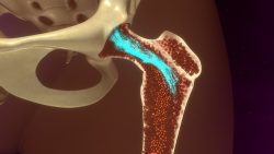
New research from the University of Cologne has found that stem cell function reduces as we age due to changes in our epigenome. Because our bones become thinner as we age, fractures and bone diseases occur more often. One reason this may happen may be impaired function of bone-marrow stem cells, which are necessary for bone maintenance integrity.

The team wanted to know why these stem cells don’t produce as much material for developing and maintaining bones as a person ages, which can cause more fat in the bone marrow to accumulate.  To find out they compared the epigenome in bones of young and old mice.

They found that the epigenome can change naturally with aging. The genes that are significant for the production of bone are especially affected.  The epigenome had differences that were from proteins called histones.  Histones combine the DNA in cells controlling access to DNA and make certain genes active or inactive. Histone modification in stem cells reduces over time which lowers activity of bone forming genes.

The research group studied the epigenome of mesenchymal stem cells. These can be found in a person’s bone marrow and can produce cells of different types such as bone, cartilage and fat cells.  This was to find out if the change in the epigenome might be a cause of higher risk to osteoporosis or bone fractures in humans. Older aged patients with osteoporosis had presented the same epigenetic changes as they had noticed in the mice.

They wondered if the epigenome stem cells could be rejuvenated. They used a nutrient solution that contained sodium acetate to treat isolated stem cells that were from mouse bone marrow. The result was the cell converting the acetate into a sequence where the enzyme attaches to histones which can promote access to genes, thus increasing their activity. This successfully rejuvenated the epigenome which improved the activity of the stem cells and led to a higher accumulation of stem cells.

They did note that treating osteoporosis with the form of sodium acetate that is a food additive is not advisable since their observation of certain cells was very specific. But stem cell therapies for treating osteoporosis are currently being undergone. It may be possible to use sodium acetate in these cases. More research is needed to determine the outcome on the whole organism to reduce possible side effects and risks.

To view the original scientific study click below:
Chromatin remodeling due to degradation of citrate carrier impairs osteogenesis of aged mesenchymal stem cells

Harvested Stem Cells for Creating Cartilage Tissue

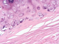
Research from the Univ. of Southampton has invented a novel way to use stem cells to generate tissue from human cartilage. This new technique could open up pathways for developing a much needed treatment for cartilage damage in people.

Cartilage serves as a shock buffer for the joints, however it is vulnerable to joint damage through trauma from falls or sports injuries or just daily wear and tear. The current surgical approach is to restore damaged cartilage regions utilizing cartilage cells but this has not been successful so far. This is due to the fact that survival of the cartilage tissue repair, which is caused by cartilage type cells at the damage site, has been shown to decrease just 5 to 10 years after the repair. There is a vital requirement for a new way to encourage long-term, robust repair through cartilage tissue implantation rather than to cartilage cells at the location of the damage.

Researchers believe they have discovered the answer to the dilemma. They generated tissue from cartilage in the lab through the success of transforming embryonic stem cells to cartilage cells. They then utilized these cells to generate 3-dimensional pieces of tissue from cartilage without using any natural or synthetic supporting substances. This is known as a cartilage engineering technique that is scaffold free. This new generated cartilage tissue produced is mechanically and structurally comparable to normal human cartilage and has the potential for a long lasting and stable repair that isn’t currently available to people.

The researchers for the study were the initial team to use this scaffold-free engineering technique to produce cartilage tissue which has been scaled up 1 mm with no adverse affects to its mechanical and structural properties. They hope that eventually following more research, the laboratory created tissue could routinely be used in surgery to heal damaged cartilage.

The research is exciting because the team’s ability to generate cartilage with properties similar to normal human cartilage will have the potential to provide vigorous tissue that has been engineered for repair of damaged cartilage.

The team thinks this tissue based approach of replacing like-for-like cartilage has the ability to form a step-by-step change improvement in the current cell based surgeries for the repair of damaged cartilage and improve the patient’s long term future and outcomes from the cartilage repairs.

To view the original scientific study click below:
A scaffold-free approach to cartilage tissue generation using human embryonic stem cells