
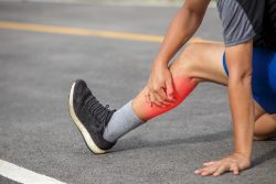
 Many people know that an injury to tendons can be difficult, lengthy and many times an incomplete process of healing. As an example, repetitive or sudden motion which can be experienced by factory workers and athletes can increase the risk of ruptures or tears in the tendons. 30% of people will experience an injury to the tendon with the highest risk in women. Additionally, people who endure this type of injury are more likely to experience a future injury at the same site or never fully recover from the injury.
Muscles which are attached to bone are tendons which are fibrous connective tissues. The tissues are soft and connected to rigid bones which makes for a difficult interface that is a very defined structure. After an injury occurs, the structure becomes obstructed and the tissues that connect the muscle to the bone change from a linear formation to one that is kinked. Excessive scarring may additionally occur which changes the tendon’s natural properties and the capacity to bear loads.
While the body is going through its natural process of healing, tendons and other cells initiate to reconstruct the primary matrix of connective parallel tissue fibers. However, this process can take weeks and even months and the resulting tendon is very often not perfect. This will result in chronic pain, weakness and a lesser quality of life.
Potential treatments for injuries to the tendons include grafts from the tendon tissue taken from donors or patients. However these have a risk of infection, necrosis or transplant rejection. Transplants that are synthetic have been tried, but biocompatibility, bio-degradation and mechanical problems have hindered these attempts.
An alternative approach is to use MSCs (mesenchymal stem cells) which are specialized cells whose pivotal role is in regeneration of tissue. At the site of the wound, they can tell the difference between various cell types. They then produce molecules that signal which will regulate cellular migration, immune response, and new formation of blood vessels which enables regeneration of tissue.
However, methods of treatment that use direct injection, systemic infusion or MSCs being genetic modified have their own problems. Infusion that is systemic does not target specifically to the site of injury. An injection that is direct requires a prohibitively large number of cells, and modification that is genetic is insufficient and will produce cells that can be hard to isolate.
Another approach has been constructing bio-material scaffolds or frameworks, and then let in MSCs and growth factors to create new tissue in the tendon. A team has used this method in developing a procedure that is showing important advances in MSC regeneration of tissue.
The team initially looked to silk fibroin which is a silk protein that is produced by the Bombyx mori silkworm. It is used in silk fabrics and additionally used in electrical and optical material and in a variety of biomedical applications such as suture materials to bone, bio-engineered ligaments, and corneal tissue. Due to its superior durability, strength, bio-degradative, and bio-compatibility aspects, silk fibroin is perfect to use in frameworks for tendons.
To enhance the ability of the framework for regeneration of tissue, the team combined silk fibroin with GelMA, a gel that is gelatin based. It retains water due to the GelMA’s controllable degradation, biocompatiblity, stiffness and the ability to promote cell growth and attachment.
The synergistic results of GelMA’s capacity for the support of formation of regenerative tissue and the structural advantages of the silk fibroin make the composite material very well suited for repair of tendons.
The team prepared mixtures of a variety of ratios of silk fibroin and GelMA (SG) and created thin nanofiber sheets. Next they tested the sheets for fiber stretchiness and structure and chose the best formulation with the optimal mechanical properties. They noticed that the silk fibroin passed on an increased permeability to the material which enhanced repair of the tissue.
The improved SG sheets were then implanted with MSCs and put through a variety of tests measuring MSC production of growth factor, differentiation and compatibility, and gene activity that triggered matrix formulation.
The MSCs that were on the SG sheets indicated a rise in proliferation and viability of cells compared to those on silk fibroin sheets that did not have the GelMA (SF). Genetic analysis indicated that pertinent gene activity in SG MSCs was greatly increased as contrasted to those on SF sheets which was shown to be decreased.
Using stained test procedures it was shown that the MSCs on the SG sheets indicated a rate higher than 80% of attachment and showed an oblong shape which is similar to cells that are attached to surfaces. This was in comparison to a 60% rate of attachment on SF and GelMA only surfaces with spherically shaped cells.
Additional tests on a growth factor that was secreted by MSCs implanted onto nanofiber sheets indicated that the growth factors produced had the best ability to repair tendon tissue that was injured.
Experiments on live rats with injuries to their Achilles tendons were also performed. MSC implanted nanofiber sheets were attached to the site of injury and the SG sheets promoted the best healing acceleration with reduced sites of injury and remodeled muscle component and the formation of densely packed, well-aligned fiber.
Remodeling of tissue for repair of tendons is very challenging to achieve. The research done has significantly advanced that achievement.
To view the original scientific study click below:
Co-Electrospun Silk Fibroin and Gelatin Methacryloyl Sheet Seeded with Mesenchymal Stem Cells for Tendon Regeneration


