
Covid-19 and Sleep Changes
 During the last 3 months of the COVID-19 pandemic, changes in sleep are just one of the many daily activities that have been impacted by this virus. A report from late March 2020 by Fitbit has shown that their sleep tracker app was showing major changes in sleep patterns for millions of Americans.
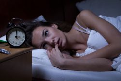
Getting a good might’s sleep is a highly important daily routine that many people don’t do. And whether it is due to child care, work, a noisy environment or other reasons, few Americans actually make sleep a priority. About 68% or 164 million Americans struggle with sleep at least once during a week. Millions of people suffered from insomnia before the pandemic, and unfortunately the pandemic has created a host of new challenges even for those people who previously had no sleep problems.
With social distancing in place along with quarantines, school closures, and working from home, profound changes to normal daily routines for people of all ages and walks of life have occurred.
Fitbit report shows that people are going to bed later but are getting more sleep in addition to a better quality of sleep. For those people whose quality of sleep has shown improvement, they have been spending more time in REM or deep sleep. Our bodies recharge while we sleep both repairing and building muscle and tissue. Levels of cortisol, our stress hormone, fall during the evening. Getting the recommended seven to nine hours of sleep per night will help boost the immunity – something that is especially important now.
Sleep is critical to both physical health and effective functioning of the immune system. It is also a key promoter of mental health and emotional wellness. It helps to beat back depressions, stress and anxiety.
From a report by the Smithsonian in late April 2020, researchers started to study other more ominous changes in sleep patterns. Sleep study centers from around the world reported increasingly vivid and strange dreams from those who had entered their second month of stay at home orders.
It was reported that a growing group are experiencing insomnia which is an inability to fall asleep which seems to be symptoms of stress due in part to the shared anxiety surrounding the COVID-19 virus. In the same report, researchers in France at The Lyon Neuroscience Research Center discovered a 35% increase in dream recall and a 15% increase in negative dreams.
For people who are not on the front lines of emergency and healthcare response, fears of the virus are projected onto threats such as bug, zombies, and shadowy figures which represent the pandemic metaphorically.
Dreams typically occur during the REM phase of sleep. Low activity and anxiety during the day can make it more difficult to get a good night’s sleep. Additionally, waking up frequently throughout the night can increase the likelihood that dreams will be remembered the next day.
REM sleep which is associated with dreaming, is believed to help us handle intense emotions and particularly negative emotions. And obviously the COVID-19 pandemic is producing a lot of anxiety and stress.
It is imperative that we let our bodies and brains rest and relax. This not only allows us to enter REM sleep and cope with stress, but a good night’s sleep also increases the production of vital neurotransmitters and hormones that our brain needs replenished every day.
The Sleep Foundation has ways we can cope with the profound changes of activity and stress that may be affecting sleep during the pandemic. Set a schedule and routine and avoid variations in daily sleep time. This includes waking up the same time every day, winding down time to relax to get ready for sleep, showering and dressing even when not leaving the house, eating meals at the same time each day, blocking of specific times for exercise and work, and avoiding naps.
We should also avoid bringing a computer to bed to do work or watch a movie in bed. It is also beneficial to frequently change bed sheets, fluff pillows and making the bed each day so it feels fresh when going to bed.
Exposure to light also plays a critical role in helping our bodies regulate sleep. Light based cues have a positive effect on circadian rhythm. If possible, spend some time outside in natural light during the day. As much as possible, open blinds and windows to let light in during the day.
And very importantly, avoid too much blue light which is produced by electronic devices. Blue light interferes with the body’s natural sleep promoting process. Excess screen time especially later in the day can have detrimental impact on sleep. It not only stimulates the brain in ways that make it difficult to wind down, but blue light from screens can suppress the natural production of melatonin which is a hormone they body makes to help us sleep.
And lastly, stay active. Continuing or starting regular daily activities has a variety of important benefits including for sleep.
To view the original scientific study click below
 Recent research has speculated that mouthwash may inhibit the spread of SARS-CoV-2 which is the virus responsible for COVID-19. The scientific review has suggested that publicly available mouthwashes may in theory, inhibit this virus.
  According to a recent most exhaustive study carried out to date, researchers have found that consuming a diet rich in fish or Omega 3 modulates the concentration of lipids which are passed on to cells through lipoproteins and can reduce the risk of cardiovascular disease. The research was carried out by Universitat Rovira I Virgili along with researchers from Harvard Medical School.
  Patients who exhibit a severe deficiency of Vitamin D are twice as likely to experience significant complications from COVID-19. Led by Northwestern University, the team conducting the research studied global data from the current COVID-19 pandemic and have discovered a strong correlation between a severe Vitamin D deficiency and mortality rates in patients with the virus.
 Both the loss of taste and smell have been anecdotally linked to a higher chance of developing the COVID-19 virus. The recent study has shown people are more than 10 times more likely to develop the virus infection than any other causes of infection.
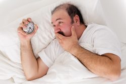 New research has found that quite the opposite is true. Previously a growing body of research has suggested that a poor quality of sleep is linked to an increased risk of obesity through deregulating appetite which results in higher caloric consumption. However, the latest study actually shows a flip side to that suggesting obesity leads to poor sleep. It isn’t the loss of sleep that leads to obesity, but instead it is excess weight that can lead to poor sleep.
  With the world in the middle of the COVID-19 pandemic, public health policies that could decrease the danger of infection and even death along with less quarantines are greatly needed. Vitamin D supplementation shows promise in reducing those risks in both the COVID-19 virus and influenza.
 According to a new study by researchers at Stanford University of Medicine, old human cells will return to a more vigorous and youthful state following being induced to briefly express a protein panel involved in embryonic development. They team also discovered that elderly mice regained their youthful strength after their existing muscle stem cells were subjected to the protein treatment rejuvenation and then transplanted back into their bodies.
 During the beginning of the COVID-19 Pandemic CDC recommends people wear face masks when in public. Due to the shortage of N95 and surgical masks, they have recommended people make their own coverings. Some materials can filter out particles much better than others, significantly affecting how well a homemade face mask may protect a person from the virus.
 A current study under leadership at the University Hospital Bonn, has concluded that a diet high in salt not only is not good for blood pressure, but also for a persons immune system. Mice who were fed a diet high in salt suffered from more serious bacterial infections. Human participants who also consumed high levels of salt also showed higher immune deficiencies.