
Preserve Bone Density As You Age
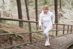 With aging, there’s a noticeable reduction in both the frequency and vigor of physical activities, contributing to deteriorating bone health. New research underscores the significance of both organized exercises and routine daily movements in preserving bone integrity. The evidence points to the advantages of integrating activities that exert stress on bones, like fast-paced walking or ascending stairs, into daily habits, underscoring their effectiveness in bone health maintenance.
Research discovered that participation in an exercise program over the course of a year enables elderly individuals (ages 70-85) to preserve or even marginally enhance the structural characteristics of their femoral neck, even amidst a decline in bone mineral density.
The subjects, who had not been active physically before, participated in a comprehensive exercise training program. Additionally, half of the participants were involved in a computer-based training program aimed at improving their information processing abilities. Physical activity levels, including the amount and intensity, were monitored using accelerometers before and after a six-month period of training. To evaluate changes in the femoral neck’s bone density and structural characteristics, dual-energy X-ray absorptiometry (DXA) scans were conducted both at the start and conclusion of the year-long exercise program.
Notably, participants who incorporated higher levels of moderate and intense activities into their regimen showed a slower reduction in bone density compared to those who engaged in activities of lower intensity or frequency.
The evidence strongly supports the integration of bone-strengthening activities, such as brisk walking and stair climbing, into daily routines as a means to counteract bone density loss. These findings advocate for a proactive approach to bone health through physical activity, underscoring its importance in the maintenance of skeletal integrity as we age.
To view the original scientific study click below:
Changes in femoral neck bone mineral density and structural strength during a 12-month multicomponent exercise intervention among older adults – Does accelerometer-measured physical activity matter?
 Recent studies have revealed an alarming increase in the prevalence of forgetfulness and confusion among working-age adults, dispelling the notion that these symptoms are merely a consequence of aging. Recent data has shown that rates of early-onset dementia and Alzheimer’s disease have unexpectedly doubled among Americans under the age of 65 between 2013 and 2017. Notably, approximately 35% of adults in the United States suffer from vitamin D deficiency. In light of these significant developments, new research has identified low levels of vitamin D as one of the 15 modifiable lifestyle factors correlated with an elevated risk of early-onset dementia.
 A significant proportion of individuals aged 65 and above in North America experience substantial muscular atrophy, scientifically known as sarcopenia, which greatly restricts their daily activities. Engaging in a consistent exercise regimen is the most effective approach to attenuate this progressive decline in strength and coordination.
  According to recent data, a significant proportion of Americans, approximately one-third, experience insomnia, leading some to self-administer sleep aids as a potential treatment for this condition. However, emerging research suggests that the benefits of a restful night’s sleep may come at a substantial cost. A striking association has been discovered between the use of sleep aids and an alarming 80% increased risk of developing dementia.
 Restricting calories has long been recognized as a means to enhance overall health and extend lifespan. However, the mechanisms through which it achieves these effects, particularly in terms of safeguarding brain health, have remained largely elusive. A recent study examining the effects of dietary restriction on aging and neurodegenerative brain diseases has yielded promising results.
 In a groundbreaking study by Yale University, researchers have discovered a direct link between reading books and living longer, healthier lives. Examining data from the renowned University of Michigan’s Health and Retirement Study, they made a remarkable finding. Individuals who read books for just 30 minutes a day can expect to add two more years to their lifespan compared to nonreaders.
 Plastic contamination in our water is a proven fact. Astonishingly, a study in 2018 unearthed an average of approximately 300 plastic particles in just one liter of water. However, this study primarily focused on microplastics, which are tiny fragments of plastic measuring less than 5 millimeters in length.
  There are better ways to protect your heart, and it starts with your nose. Researchers are uncovering the link between nasal breathing and a healthier cardiovascular system. New research has revealed that the way you breathe—nose versus mouth—can have a measurable im-pact on your heart’s well-being. Surprisingly, a recent study, though small, suggests that nasal breathing might be better for heart health, influencing key indicators of cardiovascular wellness such as blood pressure.
 Researchers have discovered that regular exercise and rest can align your body’s internal clocks, from your joints and spine to your brain. This synchronization can have incredible benefits, from boosting skeletal health to enhancing athletic performance and preventing injuries. And although this study was conducted on mice, the similarities in our cartilage and intervertebral discs lead scientists to believe that humans will experience similar results.
 A new study shows that aligning meals with natural circadian rhythms and observing a long nightly fast can greatly benefit cardiovascular health, especially in women. Our diet plays a crucial role in our overall well-being and life expectancy. The quantity, quality, and timing of our meals all have significant impacts.